

Malignant rhabdoid tumor of the kidney combined with multicystic dysplasia in a 5-year-old child
- PMID: 20436719
- PMCID: PMC2858842
- DOI: 10.3346/jkms.2010.25.5.785
Malignant rhabdoid tumor of the kidney combined with multicystic dysplasia in a 5-year-old child
Abstract
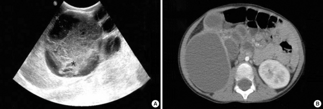
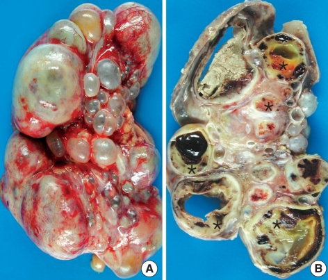
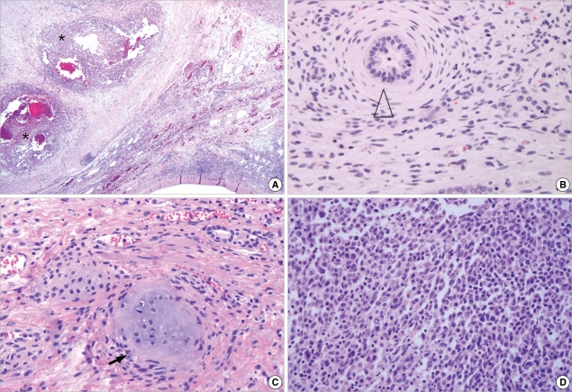
Multicystic dysplastic kidney (MCDK) is a relatively common developmental anomaly in infants and children and has a good prognosis. In contrast, a malignant rhabdoid tumor of the kidney (MRTK) is one of the most lethal neoplasms of early life. However, the presentation of such a lethal tumor combined with multicystic dysplasia has not been reported to date. In this report, we describe a case of MRTK in a 5-yr-old girl who also had multicystic dysplasia. She was previously diagnosed with MCDK at birth due to a huge palpable mass on the right side of the abdomen. The right kidney was extensively replaced by numerous grossly dilated, variable-sized cysts. Microscopically, the tumor cells show a diffusely infiltrative growth pattern, which revealed large non-cohesive, round-to-polygonal tumor cells with vesicular nuclei. Some tumor cells had eccentric nuclei and large, round, eosinophilic cytoplasmic inclusions. There were metanephrons present, with the central ureteric bud and peripheral branches surrounded by condensing mesenchyma, immature glomeruli, and metaplastic cartilage in the adjacent parenchyma. To our knowledge, this is the first combined case of the two aforementioned diseases and this case may, in fact, suggest a new disease entity.
Keywords: Childhood; Multicystic Dysplastic Kidney; Prognosis; Rhabdoid Tumor.
Figures
Similar articles
-
Rhabdoid tumour of the kidney: a diagnostic challenge and a fatal outcome.Pediatr Surg Int. 2000;16(5-6):449-50. doi: 10.1007/s003839900290. Pediatr Surg Int. 2000. PMID: 10955590
-
Antenatally diagnosed giant multicystic dysplastic kidney resected during the neonatal period.J Pediatr Surg. 2008 Nov;43(11):2118-20. doi: 10.1016/j.jpedsurg.2008.07.010. J Pediatr Surg. 2008. PMID: 18970954
-
High Activation of the AKT Pathway in Human Multicystic Renal Dysplasia.Pathobiology. 2020;87(5):302-310. doi: 10.1159/000509152. Epub 2020 Sep 14. Pathobiology. 2020. PMID: 32927453
-
Update on Multicystic Dysplastic Kidney.Curr Urol Rep. 2015 Oct;16(10):67. doi: 10.1007/s11934-015-0541-7. Curr Urol Rep. 2015. PMID: 26255066 Review.
-
Supporting Infants with Multicystic Dysplastic Kidney Disease: A Comprehensive Approach.Neonatal Netw. 2024 Oct 1;43(5):286-294. doi: 10.1891/NN-2024-0007. Neonatal Netw. 2024. PMID: 39433342 Review.
Cited by
-
Multicystic dysplastic kidney (MCDK) in the neonate: The role of the urologist.Can Urol Assoc J. 2016 Jan-Feb;10(1-2):18-24. doi: 10.5489/cuaj.3520. Can Urol Assoc J. 2016. PMID: 26977201 Free PMC article. No abstract available.
-
Imaging of Kidney Cysts and Cystic Kidney Diseases in Children: An International Working Group Consensus Statement.Radiology. 2019 Mar;290(3):769-782. doi: 10.1148/radiol.2018181243. Epub 2019 Jan 1. Radiology. 2019. PMID: 30599104 Free PMC article.
References
-
- Welch TR, Wacksman J. The changing approach to multicystic dysplastic kidney in children. J Pediatr. 2005;146:723–725. - PubMed
-
- Weeks DA, Beckwith JB, Mierau GW, Luckey DW. Rhabdoid tumor of kidney. A report of 111 cases from the national wilms' tumor study pathology center. Am J Surg Pathol. 1989;13:439–458. - PubMed
-
- Han TI, Kim MJ, Yoon HK, Chung JY, Choeh K. Rhabdoid tumour of the kidney: imaging findings. Pediatr Radiol. 2001;31:233–237. - PubMed
-
- Robson WL, Leung AK, Thomason MA. Multicystic dysplasia of the kidney. Clin Pediatr (Phila) 1995;34:32–40. - PubMed
Publication types
MeSH terms
LinkOut - more resources
Full Text Sources
Medical
