

Presacral tumors: diagnosis and management
- PMID: 20436832
- PMCID: PMC2780241
- DOI: 10.1055/s-0029-1223839
Presacral tumors: diagnosis and management
Abstract
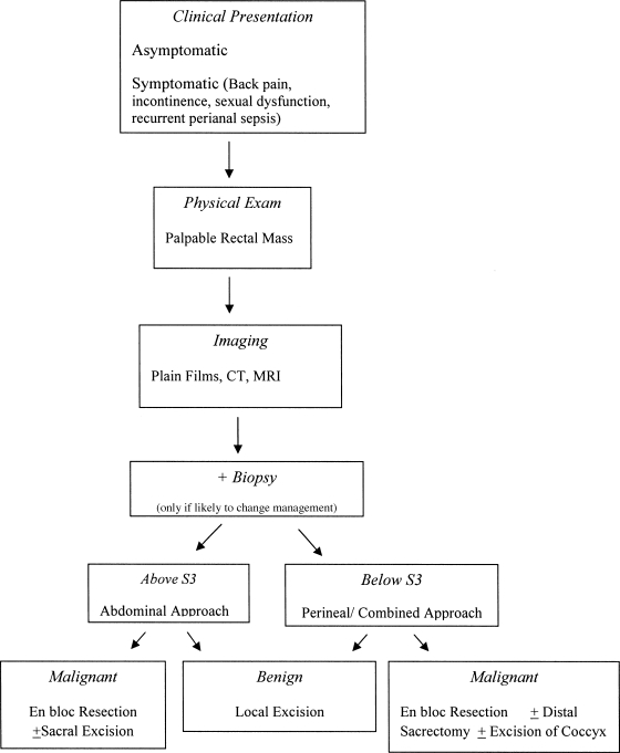
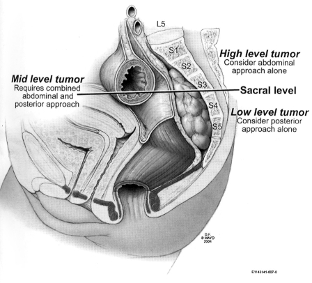
Presacral tumors are uncommon lesions that can be difficult to diagnose because of their nonspecific presenting signs and symptoms. Cross-sectional imaging is essential in evaluating these lesions to determine the optimal surgical approach and the extent of resection. Surgery is the mainstay of treatment as it establishes the diagnosis and prevents the adverse consequences associated with malignant degeneration and secondary bacterial infection. The outcomes for patients with benign presacral tumors are favorable. Although there have been substantial improvements in the prognosis of patients with malignant presacral tumors, the development of newer adjuvant therapies are likely to further improve the oncologic outcomes of malignant presacral tumors such as chordomas and sarcomas.
Keywords: Presacral tumors; diagnosis; management; prognosis; surgery.
Figures

References
-
- Hobson K G, Ghaemmaghami V, Roe J P, Goodnight J E, Khatri V P. Tumors of the retrorectal space. Dis Colon Rectum. 2005;48(10):1964–1974. - PubMed
-
- Uhlig B E, Johnson R L. Presacral tumors and cysts in adults. Dis Colon Rectum. 1975;18(7):581–589. - PubMed
-
- Gordon P H. In: Gordon PH, Nivatvongs S, editor. Principles and Practice of Surgery for the Colon, Rectum and Anus. St. Louis, MO: Quality Medical Publishing; 1999. Retrorectal tumors. pp. 427–445.
-
- Young-Fadok T M, Dozois E J. In: Yeo CJ, editor. Shackelford's Surgery of the Alimentary Tract. Philadelphia: Saunders; 2007. Retrorectal tumors. pp. 2299–2311.
-
- Dozois E J, Jacofsky D J, Dozois R R. In: Wolff BG, Fleshman JW, Beck DE, et al, editor. The ASCRS Textbook of Colon and Rectal Surgery. New York: Springer; 2007. Presacral tumors. pp. 501–514.
LinkOut - more resources
Full Text Sources
