

The relationship of retinal vessel diameter to changes in diabetic nephropathy structural variables in patients with type 1 diabetes
- PMID: 20437026
- PMCID: PMC2892559
- DOI: 10.1007/s00125-010-1763-3
The relationship of retinal vessel diameter to changes in diabetic nephropathy structural variables in patients with type 1 diabetes
Abstract
Aims/hypothesis: We examined whether retinal vessel diameter in persons with type 1 diabetes mellitus is associated with changes in subclinical anatomical and functional indicators of diabetic nephropathy.
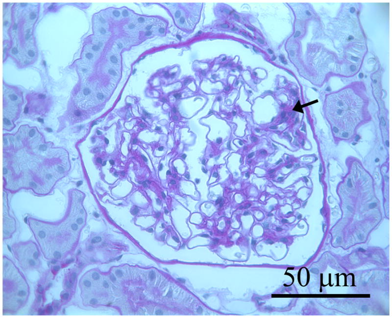
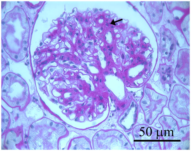
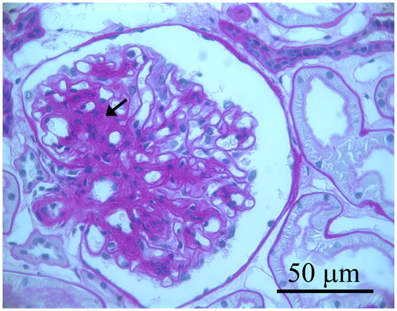
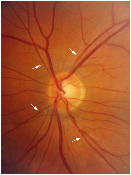
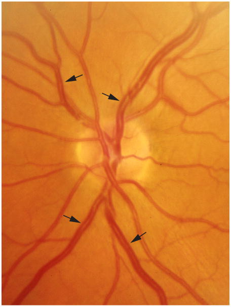
Methods: Persons with type 1 diabetes mellitus had gradable fundus photographs and renal biopsy data at baseline and 5-year follow-up (n = 234). Retinal arteriolar and venular diameters were measured at baseline and follow-up. Central retinal arteriole equivalent (CRAE) and central retinal venule equivalent (CRVE) were computed. Baseline and 5-year follow-up renal structural variables were assessed by masked electron microscopic morphometric analyses from percutaneous renal biopsy specimens. Variables assessed included: mesangial fractional volume, glomerular basement membrane width, mesangial matrix fractional volume and glomerular basement membrane width composite glomerulopathy index.
Results: While controlling for other covariates, baseline CRAE was positively associated with change in the glomerulopathy index over the 5-year period. Change in CRAE was inversely related to a change in mesangial matrix fractional volume and abnormal mesangial matrix fractional volume, while change in CRVE was directly related to change in the volume fraction of cortex that was interstitium [Vv((Int/cortex))] over the 5-year period. Baseline CRAE or CRVE or changes in these diameters were not related to changes in other anatomical or functional renal endpoints.
Conclusions/interpretation: Independently of other factors, baseline CRAE correlated with changes in glomerulopathy index, a composite measure of extracellular matrix accumulation in the mesangium and glomerular basement membrane. A narrowing of the CRAE was related to mesangial matrix accumulation. Changes in CRVE were related to changes in Vv((Int/cortex),) a measure of interstitial expansion in persons with type 1 diabetes mellitus.
Conflict of interest statement
Dr. Zinman, lecture fees, consulting fees, and research grants from Merck; and Dr. Klein reports being an advisory board member for AstraZeneca (through the DIRECT study), Pfizer, Lilly, and Novartis.
No other dualities of interest relevant to this article were reported.
Figures
References
-
- National Institutes of Health, National Institute of Diabetes and Digestive and Kidney Diseases. U.S. Renal Data System, USRDS 2006 annual data report: atlas of end-stage renal disease in the United States. Bethesda, MD: National Institutes of Health, National Institute of Diabetes and Digestive and Kidney Diseases; 2006.
-
- Reddi AS, Camerini-Davalos RA. Diabetic nephropathy. An update. Arch Intern Med. 1990;150:31–43. - PubMed
-
- Raptis AE, Viberti G. Pathogenesis of diabetic nephropathy. Exp Clin Endocrinol Diabetes. 2001;109(Suppl 2):S424–S437. - PubMed
-
- Jawa A, Kcomt J, Fonseca VA. Diabetic nephropathy and retinopathy. Med Clin North Am. 2004;88:1001–36. xi. - PubMed
-
- Gross JL, de Azevedo MJ, Silveiro SP, Canani LH, Caramori ML, Zelmanovitz T. Diabetic nephropathy: diagnosis, prevention, and treatment. Diabetes Care. 2005;28:164–176. - PubMed
Publication types
MeSH terms
Grants and funding
LinkOut - more resources
Full Text Sources
Medical
