

Growth, cell division and sporulation in mycobacteria
- PMID: 20437098
- PMCID: PMC2906719
- DOI: 10.1007/s10482-010-9446-0
Growth, cell division and sporulation in mycobacteria
Abstract
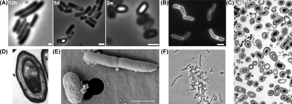
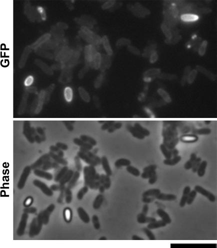
Bacteria have the ability to adapt to different growth conditions and to survive in various environments. They have also the capacity to enter into dormant states and some bacteria form spores when exposed to stresses such as starvation and oxygen deprivation. Sporulation has been demonstrated in a number of different bacteria but Mycobacterium spp. have been considered to be non-sporulating bacteria. We recently provided evidence that Mycobacterium marinum and likely also Mycobacterium bovis bacillus Calmette-Guérin can form spores. Mycobacterial spores were detected in old cultures and our findings suggest that sporulation might be an adaptation of lifestyle for mycobacteria under stress. Here we will discuss our current understanding of growth, cell division, and sporulation in mycobacteria.
Figures
Similar articles
-
Sporulation in mycobacteria.Proc Natl Acad Sci U S A. 2009 Jun 30;106(26):10781-6. doi: 10.1073/pnas.0904104106. Epub 2009 Jun 16. Proc Natl Acad Sci U S A. 2009. PMID: 19541637 Free PMC article.
-
Mycobacterium versus Streptomyces--we are different, we are the same.Curr Opin Microbiol. 2009 Dec;12(6):699-707. doi: 10.1016/j.mib.2009.10.003. Epub 2009 Oct 31. Curr Opin Microbiol. 2009. PMID: 19880345 Review.
-
Do mycobacteria produce endospores?Proc Natl Acad Sci U S A. 2010 Jan 12;107(2):878-81. doi: 10.1073/pnas.0911299107. Epub 2009 Dec 22. Proc Natl Acad Sci U S A. 2010. PMID: 20080769 Free PMC article.
-
Asymmetric cell division during Bacillus subtilis sporulation.Future Microbiol. 2019 Mar;14:353-363. doi: 10.2217/fmb-2018-0338. Epub 2019 Mar 11. Future Microbiol. 2019. PMID: 30855188
-
Bacterial growth and cell division: a mycobacterial perspective.Microbiol Mol Biol Rev. 2008 Mar;72(1):126-56, table of contents. doi: 10.1128/MMBR.00028-07. Microbiol Mol Biol Rev. 2008. PMID: 18322037 Free PMC article. Review.
Cited by
-
Age-Dependent Pleomorphism in Mycobacterium monacense Cultures.Microorganisms. 2025 Feb 20;13(3):475. doi: 10.3390/microorganisms13030475. Microorganisms. 2025. PMID: 40142368 Free PMC article.
-
Mycobacterial Growth.Cold Spring Harb Perspect Med. 2015 May 8;5(10):a021097. doi: 10.1101/cshperspect.a021097. Cold Spring Harb Perspect Med. 2015. PMID: 25957314 Free PMC article. Review.
-
Comparative Sigma Factor-mRNA Levels in Mycobacterium marinum under Stress Conditions and during Host Infection.PLoS One. 2015 Oct 7;10(10):e0139823. doi: 10.1371/journal.pone.0139823. eCollection 2015. PLoS One. 2015. PMID: 26445268 Free PMC article.
-
Arrayed CRISPRi and quantitative imaging describe the morphotypic landscape of essential mycobacterial genes.Elife. 2020 Nov 6;9:e60083. doi: 10.7554/eLife.60083. Elife. 2020. PMID: 33155979 Free PMC article.
-
Cell division site placement and asymmetric growth in mycobacteria.PLoS One. 2012;7(9):e44582. doi: 10.1371/journal.pone.0044582. Epub 2012 Sep 10. PLoS One. 2012. PMID: 22970255 Free PMC article.
References
-
- Ara I, Kudo T. Three novel species of the genus Catellatospora, Catellatospora chokoriensis sp. nov., Catellatospora coxensis sp. nov. and Catellatospora bangladeshensis sp. nov., and transfer of Catellatospora citrea subsp. methionotrophica Asano and Kawamoto 1988 to Catellatospora methionotrophica sp. nov., comb. nov. Int J Syst Evol Microbiol. 2006;56:393–400. doi: 10.1099/ijs.0.63862-0. - DOI - PubMed
-
- Asano K, Kawamoto I. Catellatospora, a new genus of the Actinomycetales. Int J Syst Bacteriol. 1986;36:512–517. doi: 10.1099/00207713-36-4-512. - DOI
-
- Ben-Yehuda S, Rudner DZ, Losick R. Assembly of the SpoIIIE DNA translocase depends on chromosome trapping in Bacillus subtilis. Curr Biol. 2003;13:2196–2200. - PubMed
Publication types
MeSH terms
LinkOut - more resources
Full Text Sources
Other Literature Sources
Molecular Biology Databases
Miscellaneous
