. 2010 Sep;19(9):1468-78.
doi: 10.1007/s00586-010-1383-0. Epub 2010 May 1.

The influence of torsion on disc herniation when combined with flexion

Affiliations

The influence of torsion on disc herniation when combined with flexion

Samuel P Veres et al. Eur Spine J. 2010 Sep.

Abstract

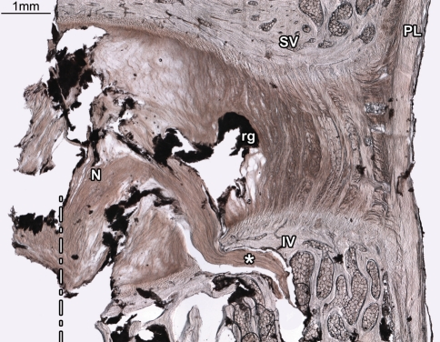
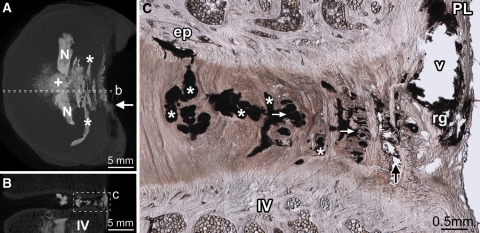
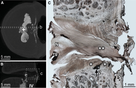
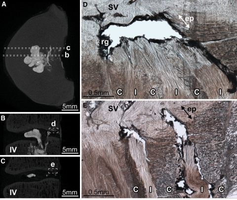
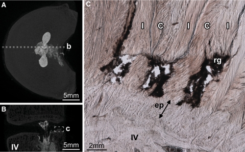
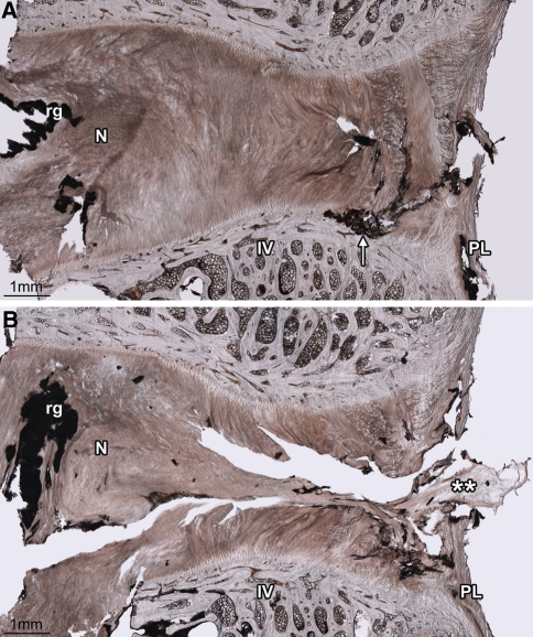
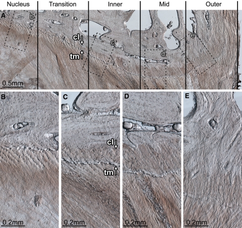
The role of torsion in the mechanical derangement of intervertebral discs remains largely undefined. The current study sought to investigate if torsion, when applied in combination with flexion, affects the internal failure mechanics of the disc wall when exposed to high nuclear pressure. Thirty ovine lumbar motion segments were each positioned in 2 degrees axial rotation plus 7 degrees flexion. Whilst maintained in this posture, the nucleus of each segment was gradually injected with a viscous radio-opaque gel, via an injection screw placed longitudinally within the inferior vertebra, until failure occurred. Segments were then inspected using micro-CT and optical microscopy in tandem. Five motion segments failed to pressurize correctly. Of the remaining 25 successfully tested motion segments, 17 suffered vertebral endplate rupture and 8 suffered disc failure. Disc failure occurred in mature motion segments significantly more often than immature segments. The most common mode of disc failure was a central posterior radial tear involving a systematic annulus-endplate-annulus failure pattern. The endplate portion of these radial tears often propagated contralateral to the direction of applied axial rotation, and, at the lateral margin, only those fibres inclined in the direction of the applied torque were affected. Apart from the 2 degrees of applied axial rotation, the methods employed in this study replicated those used in a previously published study. Consequently, the different outcome obtained in this study can be directly attributed to the applied axial rotation. These inter-study differences show that when combined with flexion, torsion markedly reduces the nuclear pressure required to form clinically relevant radial tears that involve cartilaginous endplate failure. Conversely, torsion appears to increase the disc wall's resistance to radial tears that do not involve cartilaginous endplate failure, effectively halving the disc wall's overall risk of rupture.


Figures

Fig. 1
Fig. 1
Rupture of the inferior vertebral endplate was the most common mode of failure amongst tested motion segments. In this disc (2-L56) the inferior vertebral endplate has ruptured near the nucleus/annulus transition zone, allowing nuclear material to enter the inferior vertebral body (asterisks). The dashed line marks the centre of the hole through which the injection screw was inserted. The injected contrast gel (rg) has reached the inner posterior annulus, but has failed to penetrate the disc radially beyond this point, leaving the mid and outer portions of the disc intact. N nucleus, SV superior vertebra, IV inferior vertebra, PL posterior longitudinal ligament
Fig. 2
Fig. 2
Diffuse rupture of the posterior annulus. Disc 8-L56 is shown. Dashed lines in the axial (A) and sagittal (B) micro-CT MIP images show the location of the micrograph (C). From the point of injection (+ in A), contrast gel has spread laterally filling the nuclear lobes (N in A). After penetrating the inner posterior annulus, the gel has spread circumferentially, flowing within the inner and mid-annular lamellae (* in A and C). Two short radial tears, connecting some of the circumferential tears, are visible in C (white arrows). Upon reaching the outer annulus, gel has flowed primarily between lamellae (black arrow in C). In both discs that suffered diffuse ruptures, the contrast gel failed to rupture the posterior longitudinal ligament (PL in C), leaving a large void (v in C) outlined in residual gel (rg in C) as it tracked circumferentially to its point of extrusion at the ligament’s left lateral margin (white arrow in A). IV inferior vertebra, ep tear of the cartilaginous endplate
Fig. 3
Fig. 3
Radial annular-endplate tear of the central posterior disc wall was the most frequent cause of disc failure. Disc 3-L34 is shown. The axial (A) and sagittal (B) micro-CT MIP images show the location of the micrograph (C). Radial annular-endplate tears were characterized by two regions of annular failure (* in C), separated by an endplate tear adjacent to the mid-annulus (arrow2 in C) In this disc, the tear contains a large volume of nuclear material (** in C), which has severed the posterior longitudinal ligament forming a transligamentous nuclear extrusion. Prior to rupture, the separated regions marked by arrows 1 and 2 in C would have been connected. N nucleus, rg residual gel, IV inferior vertebra, PL posterior longitudinal ligament
Fig. 4
Fig. 4
The variation in endplate tear morphology with circumferential location in radial annular-endplate tears. Disc 7-L12 is shown. The axial (A) and sagittal (B, C) micro-CT MIP images show the location of the micrographs (D, E). The endplate portion of the radial annular-endplate tears spanned between 2.5 and 5.9 mm circumferentially, most often extending to the right from the discs’ midline (Table 2). At the central posterior location tears always occurred at the cartilaginous/vertebral endplate junction and extended a radial distance of approximately 1 mm, disrupting the anchorage of both in-plane (I) and cross-sectioned (C) lamellae (D). The right mediolateral aspect of the endplate tears were often morphologically distinct; only fibres inclined in the direction of the applied axial rotation were affected (I vs. C in E). IV inferior vertebra, SV superior vertebra, rg residual gel. Arrows marked ‘ep’ indicate the thickness of the cartilaginous endplate
Fig. 5
Fig. 5
The axial (A) and sagittal (B) micro-CT MIP images show the location of the micrograph (C). Whilst the endplate portion of radial annular-endplate tears usually occurred at the cartilaginous/vertebral endplate junction, in two discs the right mediolateral portion of the endplate tear occurred at the annulus/endplate junction (disc 5-L12 is shown). In both discs, only fibres inclined in the direction of the applied axial rotation were ruptured, in this case those of the in-plane lamellae (I), but not the cross-sectioned lamellae (C). IV inferior vertebra, rg residual gel. The arrow marked ‘ep’ indicates the thickness of the cartilaginous endplate
Fig. 6
Fig. 6
The unique case of disc 8-L12, shown here, highlights that whilst torsion tended to direct endplate tears contralaterally, the central posterior portion of the disc wall was most vulnerable to radial tearing. The two cryosections shown lie approximately 1.5 mm apart in the same plane. A This right mediolateral cryosection shows an endplate tear adjacent to the mid-annulus (arrow). B This central posterior cryosection shows that nuclear material (**) has bisected the annulus, forming a radial mid-axial tear resulting in a transligamentous nuclear extrusion. No form of endplate disruption is present in this cryosection. N nucleus, IV inferior vertebra, PL posterior longitudinal ligament, rg residual gel
Fig. 7
Fig. 7
Endplate morphology and disruption susceptibility. The disc wall can be divided into three morphologically distinct zones: inner, mid and outer. Between the nucleus and inner zone exists the transition zone. The transition zone contains fibres that have a well-defined superior–inferior orientation, but are not organized into distinct lamellae. Inner zone (c): the inner zone begins at the first well-defined lamella. Whilst no calcification of the cartilaginous endplate is present adjacent to the nucleus (B), adjacent to the transition and inner zones both calcified and uncalcified cartilaginous endplate exist (C). The boundary between the calcified and uncalcified cartilage, the tidemark, is indicated by arrow tm in C. The boundary between the calcified cartilage and the vertebral endplate, the cement line, is indicated by arrow cl in C. The ratio of calcified to uncalcified cartilage increases with increasing radial distance away from the nucleus. Mid-zone (d): the cartilaginous endplate is fully calcified. The cement line is well defined. There appears to be little to no connection between the annular fibres penetrating the cartilaginous endplate and the vertebral endplate. It is the endplate in this zone that is always implicated in radial annular-endplate tears and appears to be the most prone to mechanical disruption (Figs. 3C, 4D). Outer zone (e): the cartilaginous endplate is fully calcified. The cement line is less prominent. Annular fibres penetrating the cartilaginous endplate appear to integrate with the vertebral endplate forming a robust disc/vertebra connection. The tissue section shown is from the mediolateral posterior aspect of a ewe’s L56 disc (vertebral growth plates unfused). Note that the posterior longitudinal ligament has been removed; if present it would exist to the right of the outer zone

References

    1. Farfan HF. Effects of torsion on the intervertebral joints. Can J Surg. 1969;12:336–341. - PubMed
    1. Farfan HF, Cossette JW, Robertson GH, Wells RV, Kraus H. The effects of torsion on the lumbar intervertebral joints: the role of torsion in the production of disc degeneration. J Bone Joint Surg Am. 1970;52:468–497. - PubMed
    1. Adams MA, Hutton WC. The relevance of torsion to the mechanical derangement of the lumbar spine. Spine. 1981;6:241–248. doi: 10.1097/00007632-198105000-00006. - DOI - PubMed
    1. Gunzburg R, Hutton W, Fraser R. Axial rotation of the lumbar spine and the effect of flexion. An in vitro and in vivo biomechanical study. Spine. 1991;16:22–28. doi: 10.1097/00007632-199101000-00004. - DOI - PubMed
    1. Ochia RS, Inoue N, Renner SM, Lorenz EP, Lim TH, Andersson GB, An HS. Three-dimensional in vivo measurement of lumbar spine segmental motion. Spine. 2006;31:2073–2078. doi: 10.1097/01.brs.0000231435.55842.9e. - DOI - PubMed

Publication types