. 2010 Apr;67(4):488-97.
doi: 10.1002/ana.21919.

Endothelial progenitor cell transplantation improves long-term stroke outcome in mice

Affiliations

Endothelial progenitor cell transplantation improves long-term stroke outcome in mice

Yongfeng Fan et al. Ann Neurol. 2010 Apr.

Abstract

Objective: Endothelial progenitor cells (EPCs) play an important role in tissue repairing and regeneration in ischemic organs, including the brain. However, the cause of EPC migration and the function of EPCs after ischemia are unclear. In this study, we demonstrated the effects of EPCs on ischemic brain injury in a mouse model of transient middle cerebral artery occlusion (tMCAO).

Methods: Circulating human EPCs were characterized with immunofluorescent staining and flow cytometry. EPCs (1 x 10(6)) were injected into nude mice after 1 hour of tMCAO. Histological analysis and behavioral tests were performed from day 0 to 28 days after tMCAO.

Results: EPCs were detected in ischemic brain regions 24 hours after tMCAO. EPC transplantation significantly reduced ischemic infarct volume at 3 days after tMCAO compared with control animals (p < 0.05). CXCR4 was expressed in the majority of EPCs, and stromal-derived factor-1 (SDF-1) induced EPC migration, which was blocked by pretreated EPCs with AMD3100 in vitro. SDF-1 was upregulated in ischemic brain. Compared with control animals, injecting AMD3100-pretreated EPCs resulted in a larger infarct volume 3 days after tMCAO, suggesting that SDF-1-mediated signaling was involved in EPC-mediated neuroprotection. In addition, EPC transplantation reduced mouse cortex atrophy 4 weeks after tMCAO and improved neurobehavioral outcomes (p < 0.05). EPC injection potently increased angiogenesis in the peri-infarction area (p < 0.05).

Interpretation: We conclude that systemic delivery of EPCs protects the brain against ischemic injury, promotes neurovascular repair, and improves long-term neurobehavioral outcomes. Our data suggest that SDF-1-mediated signaling plays a critical role in EPC-mediated neuroprotection.


Figures

Fig 1
Fig 1. EPC culture and identification
A. Mononuclear cells isolated from human peripheral blood were plated in fibronectin-coated wells at 5×10⁵ cells /cm2 (a). After 5 days culture, the spindle-like attached cells that supposed to be EPCs (b) were collected for use (Bar=50 µm). B. The putative EPCs were able to take up DiI-labeled acetylated low density lipoprotein (Ac-LDL, a) and bind the endothelial specific lectin FITC-UEA-1 (b), which were colocalized in more than 95% cells (c) (Bar=50µm). C. Immunofluorescent staining showed the putative EPCs were positive for endothelial cell markers KDR (a), VE-Cadherin (b), vWF (c) and Tie-2 (d) (Bar=50µm). D. The cultured cells were further confirmed by flow cytometry analysis after being labeled with different markers (d, e, f, j, k, l) and fluorescent labeled IgG with the same isotypes for negative control (a, b, c, g, h, i). The results showed that ~2% cells expressed the hematopoietic cell marker CD34-PE (d) and the majority of cells expressed endothelial markers KDR (66–88%, d and e), VE-Cadherin (78%, f), CD-31 (94%, j) and monocyte markers CD11b (78%, k) and CD14 (82%, l).
Fig 2
Fig 2. Intravenously delivered EPCs home to ischemic brain
A. Photomicrographs show DiI labeled EPCs migrated across microvessel walls at 24 hours after injection (b, c) and into ischemic area after 72 hours of injection (e). Inserted boxes in each picture show the large magnification of cells. In the control group, DiI labeled HBEC did not show at 24 hours after injection (a) and few cells were observed in the ischemic zone (d). DAPI was used for the counterstaining. B. The figure shows the ischemic area and the ischemic boundary area. C. The bar graph shows that a great number of DiI-EPC homed into ischemic brains compared with DiI-HBEC 72 hours after injection i.v. Data are mean±SD, n=6 in each group. *, p<0.01, the DiI-EPC number vs. DiI-HBEC number in ischemic brain. Bar=50 µm.
Fig 3
Fig 3. EPC transplantation decreased acute ischemic infarction
A. Nissl’s staining indicates that EPCs-treated mice showed much smaller infarct area than saline-treated group. The mice were subjected to 1 hour of MCAO followed with reperfusion and EPC transplantation or equal volume of saline as a control through left jugular vein. After 3 days of reperfusion, the ischemic brains were collected and brain sections were prepared. B. Bar graph of quantitative analysis showed that systemic administration of EPC significantly reduced the infarct volume 3 days after tMCAO compared with control group. Data are mean±SD, n=6 in each group. *, p<0.05, EPC treated mice vs. saline treated mice.
Fig 4
Fig 4. EPC expressed CXCR4 and SDF-1 induced EPC migration in vitro
A. The photomicrographs show the expression of CXCR4, the receptor of SDF-1 (Red) on adult endothelial cells HBECs (a) and EPCs (b), with the counterstaining of DAPI (blue). Few adult endothelial cells expressed CXCR4 while the majority of EPCs did. Bar=50 µm. B. the scatter graph showed CXCR4 expression on EPCs detected with the flowcytometry. (a), control staining with IgG with the same isotope as CXCR4 antibody. (b), more than 80% of cultured EPCs expressed CXCR4. C. Bar graphs show that SDF-1 induced EPC migration assayed with the Boyden chamber system. After 18 hours of SDF-1 stimulation, cells cross the porous filter were counted and data are presented as fold increase in migrating cells relative to the control medium. (a) a large number of EPCs migrated across the porous filter compared with the control, while pretreatment with 5ug/ml of AMD3100 significantly inhibited SDF-1-induced EPC migration (* P<0.01, SDF-1 treated group vs. PBS treated control; **P<0.01, AMD3100 treated EPC vs. non-AMD 3100 treated group). (b) the bar graph shows hat even 1ng/ml of SDF-1 significantly increased EPC migration (*P<0.05, SDF-1-treated groups vs. PBS control group); (c) the bar graph shows as low as 1ug/ml of AMD3100 significantly inhibited the SDF-1-induced EPC migration (*P<0.05, AMD3100-treated groups vs. non-treated group). All the results are expressed as mean±SD and each panel represents the results of at least three independent experiments performed in triplicate.
Fig 5
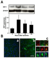
Fig 5. SDF-1 expression was up-regulated in mouse brain subjected to tMCAO following reperfusion
A. The western blot results show SDF-1 expression in ischemic brains, with β-actin as an internal control (upper panel). SDF-1 expression significantly increased at 1 day, 3 days and 7 days, with a peak at 1 day after tMCAO (lower panel). Data are mean±SD, n= 6. * P<0.05, SDF-1 expression in the brains of tMCAO mice vs. saline treated mice. B. The photomicrographs show the immunofluorescent staining of SDF-1 (green) in (a) non-ischemic brains and (b) brains subjected to 1 hour of tMCAO with 24 hours of reperfusion. Double staining (c) of SDF-1 (green) with CD31 (red, upper panel) or GFAP (red, middle panel) showed that SDF-1 was expressed in endothelial cells and astrocytes. Bar=50 µm. Based on the cell morphology, SDF-1 was also expressed in neurons (c, low panel).Bar=20 µm.
Fig 6
Fig 6. AMD3100-treated EPCs abolished cerebral protection against ischemic injury
A. Nissl’s staining showed that the infarct area of mice with AMD3100 treated-EPC was much bigger than that treated with EPCs. The mice were subjected to 1 hour of MCAO followed with reperfusion and transplantation of EPCs or 5ug/ml of AMD3100-treated EPCs through left jugular vein. After 3 days of reperfusion, the ischemic brains were collected and brain sections were prepared. B. Bar graph showed that the brain infarction volume of mice with AMD3100-treated EPCs significantly increased compared with the non-treated EPC group. Data are mean±SD, n= 6. *P<0.05, AMD3100-treated EPC group vs. non-treated-EPC group.
Fig 7
Fig 7. EPC transplantation reduced mouse cortical atrophy and improved neurobehavioral outcomes
A. EPC-treated mice showed reduced cortical atrophy after tMCAO/reperfusion. The mice were subjected to 1 hour of MCAO followed with reperfusion and EPC transplantation. Saline and HBEC were used as controls. The ischemic brains were collected 4 weeks after tMCAO. (a). Nissl’s staining showed much smaller atrophy in the brains of EPC-treated group than that of saline or HBEC-treated group. (b) Quantitative analysis showed that systemic administration of EPC significantly reduced the brain atrophy 4 weeks after tMCAO compared with saline or HBEC-treated group (b). Data are mean±SD, n=6 in each group. *, p<0.05, EPC treated mice vs. saline or HBEC treated mice. B. EPC-treated mice showed improved neurobehavioral outcome. All mice were trained for 3 days before tMCAO with 3 consecutive trials to generate stable baseline values. Neurobehavioral functions of mice were tested at 1 day, 7 days, 14 days, and 28 days after tMCAO. Line graphs show that EPC-treated mice showed significantly improved performance in crossing the beam walk (a, beam test), staying on the rotor-rod (b, rotor-rod test), and turn preferentially towards non-impaired ipsilateral side (c, corner test) compared with mice treated with saline or HBEC but there was no significant difference between saline and HBEC-treated mice 4 weeks after tMCAO. Data are mean±SD, n=6 in each group. *, p<0.05, EPC treated mice vs. saline or HBEC-treated mice.
Fig 8
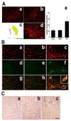
Fig 8. Intravenously delivered EPCs promoted local angiogenesis in ischemic brain
A. CD31 immunostaining shows microvessels in mouse brain treated with saline (a), HBEC (b) and EPC (d) 4 weeks after 1 hour of tMCAO. The figure (c) shows the ischemic area and the ischemic boundary area. The bar graph (e) shows that the number of microvessels in EPC-treated mice was significantly increased compared with that in saline or HBEC-treated mice. Data are mean±SD, n=6 in each group. *, p<0.05, the microvessel number in EPC-treated mice vs. that of HBEC or saline-treated mice. Bar=100 µm. B. Photomicrographs show CD31 (red, a, b, c) and BrdU (green, d, e, f) double staining in ischemic brain of saline (a, d, g), HBEC (b, e, h) and EPC (c, f, i)-treated mice. In EPC treated group, increased BrdU (+) cells were localized in endothelial cells (yellow, g, h, i), suggesting EPC transplantation promoted new vessel formation in ischemic brain. Bar=50 µm. Inserted boxes in each picture show the large magnification of cells. C. Photomicrographs human CD34-positive microvessels (arrow heads) in ischemic boundary areas of mouse brain. 4 weeks after tMCAO, anti-human CD34 antibody was used to track the transplanted human EPCs in mouse brain. The human CD34-positive microvessels were not observed in saline control group (a) or the adult HBEC-treated group (b), but formed in EPC transplanted mouse brains (c). Bar=100 µm.

References

    1. Miraglia S, Godfrey W, Yin AH, et al. A novel five-transmembrane hematopoietic stem cell antigen: isolation, characterization, and molecular cloning. Blood. 1997;90:5013–5021. - PubMed
    1. Quirici N, Soligo D, Caneva L, et al. Differentiation and expansion of endothelial cells from human bone marrow CD133(+) cells. Br J Haematol. 2001;115:186–194. - PubMed
    1. Asahara T, Murohara T, Sullivan A, et al. Isolation of putative progenitor endothelial cells for angiogenesis. Science. 1997;275:964–967. - PubMed
    1. Werner N, Kosiol S, Schiegl T, et al. Circulating endothelial progenitor cells and cardiovascular outcomes. N Engl J Med. 2005;353:999–1007. - PubMed
    1. Shintani S, Murohara T, Ikeda H, et al. Mobilization of endothelial progenitor cells in patients with acute myocardial infarction. Circulation. 2001;103:2776–2779. - PubMed

Publication types

MeSH terms