Comparative Study
. 2010 Apr;67(4):516-25.
doi: 10.1002/ana.21923.

Developmental and degenerative features in a complicated spastic paraplegia

Affiliations
Free PMC article
Comparative Study

Developmental and degenerative features in a complicated spastic paraplegia

M Chiara Manzini et al. Ann Neurol. 2010 Apr.
Free PMC article

Abstract

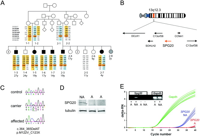
Objective: We sought to explore the genetic and molecular causes of Troyer syndrome, one of several complicated hereditary spastic paraplegias (HSPs). Troyer syndrome had been thought to be restricted to the Amish; however, we identified 2 Omani families with HSP, short stature, dysarthria and developmental delay-core features of Troyer syndrome-and a novel mutation in the SPG20 gene, which is also mutated in the Amish. In addition, we analyzed SPG20 expression throughout development to infer how disruption of this gene might generate the constellation of developmental and degenerative Troyer syndrome phenotypes.

Methods: Clinical characterization of 2 non-Amish families with Troyer syndrome was followed by linkage and sequencing analysis. Quantitative polymerase chain reaction and in situ hybridization analysis of SPG20 expression were carried out in embryonic and adult human and mouse tissue.

Results: Two Omani families carrying a novel SPG20 mutation displayed clinical features remarkably similar to the Amish patients with Troyer syndrome. SPG20 mRNA is expressed broadly but at low relative levels in the adult brain; however, it is robustly and specifically expressed in the limbs, face, and brain during early morphogenesis.

Interpretation: Null mutations in SPG20 cause Troyer syndrome, a specific clinical entity with developmental and degenerative features. Maximal expression of SPG20 in the limb buds and forebrain during embryogenesis may explain the developmental origin of the skeletal and cognitive defects observed in this disorder.


Figures

FIGURE 1
FIGURE 1
Affected individuals carry a homozygous null mutation in the SPG20 gene. (A) Pedigree of 2 related Omani families affected with short stature, spasticity, dysarthria, and developmental delay. Affected individuals are in black and unaffected in white. The status of individual 2-8 (in gray) could not be determined, because she is too young to properly assess neurological symptoms. All numbered individuals were examined, but genomic DNA was only collected from individuals for whom microsatellite analysis is shown. Microsatellite analysis revealed common maternal (in yellow) and paternal (in blue) haplotypes in all affected. Microsatellite markers in the linkage region identified by the single nucleotide polymorphism analysis are in bold. One homozygous marker (D13S219) is common to all affected individuals (highlighted in orange). (B) The homozygous region contains 6 genes, including SPG20 (in orange). (C) The affected individuals carried a homozygous 2bp deletion in SPG20, which was present in heterozygosity in the carriers. (D) Western blot analysis of patient cell lines showed that full-length SPG20 protein is missing in the affected individuals (A) compared with a nonaffected noncarrier sibling (NA). (E) Quantitative polymerase chain reaction analysis of cDNA from the patient cell lines indicated that SPG20 mRNA is not present in the affected individual. deltaRN = signal magnitude expressed by the difference in the normalized reporter (RN) values.
FIGURE 2
FIGURE 2
Morphological and radiological features of Omani individuals with Troyer syndrome. Comparison between (A, C) an unaffected and (B, D) an affected individual revealed (A, B) relative hypertelorism and (C, D) pronounced overbite. Examples of skeletal anomalies in the extremities: brachydactyly (short digits; E, F), camptodactyly (flexed digit; asterisks in E, F), hammer toes (asterisks in G, H), and clinodactyly (curved digit; arrowheads in G, H). Brain magnetic resonance imaging shows atrophy of the cerebellar vermis (arrowhead in I) and white matter hyperintensity in T2-weighted images, which is more prominent posteriorly (arrowheads in J).
FIGURE 3
FIGURE 3
Quantitative polymerase chain reaction analysis of SPG20/Spg20 expression in human and mouse tissues. (A) There is modest variation in SPG20 local expression in the adult human brain when distinct regions are compared to whole brain samples. (B) Human SPG20 expression is substantially lower than that of the brain-specific gene synaptophysin (SYP) across all brain regions with the exception of the amygdala. (C) A similar profile of regionally variable Spg20 expression, with some modest differences, is seen in the mouse brain. (D) Lower Spg20 expression is observed when compared with Syp. (E) Spg20 is expressed in several murine tissues beside the brain. (F) Spg20 is developmentally regulated in the mouse embryo with highest expression levels in the whole embryo at E10.5. ΔCT =; Bas Gang = basal ganglia; DeltaCT = signal magnitude expressed by the difference in CT values. Hippoc = hippocampus; Mesen = mesenchyme; Cerebel = cerebellum; Sp Cord = spinal cord; Sk Muscle = skeletal muscle; Rel. = relative.
FIGURE 4
FIGURE 4
In situ analysis of Spg20 expression in mice. (A–J) Spg20 expression in the adult brain is modest and focal. (E) Cortical expression is moderate and not layer-specific. Relatively higher expression is observed in (B) the hippocampus, (C) glial cells in the corpus callosum, (D) entorhinal cortex, (F, G) habenular complex, (H) cerebellum, (I) facial nucleus (fN), and (J) spinal cord. (K) Fetal expression is also modest and distributed in the brain, with higher expression in (L) the lens placode (le), (M) the cochlear epithelium, and (N) condensing mesenchyme at sites of myogenic and cartilage formation (asterisks). (O) Early embryonic expression (embryonic day 10.5) is highly patterned and enhanced in the forebrain (fb), frontonasal mass (fnm), maxilla (mx), branchial arches (ba1, ba2), heart (h), and limb buds (flb). (P) The antisense control is completely unlabeled. (Q) Selective, focally elevated expression in the fnm/fb, flb, and ba1/ba2 is confirmed by qPCR after microdissection compared to the heart (h) and whole embryo (Wh). CA1 =; CA2 =; CA3 =; CC = corpus callosum; m = molecular layer; P = Purkinje cells layer; g = granule cell layer; dh = dorsal horn; vh = ventral horn; Ctx = cortex; LGE = lateral ganglionic eminence; MGE = medial ganglionic eminence; r = retina.

References

    1. Harding AE. Classification of the hereditary ataxias and paraplegias. Lancet. 1983;1:1151–1155. - PubMed
    1. Depienne C, Stevanin G, Brice A, Durr A. Hereditary spastic paraplegias: an update. Curr Opin Neurol. 2007;20:674–680. - PubMed
    1. Salinas S, Proukakis C, Crosby A, Warner TT. Hereditary spastic paraplegia: clinical features and pathogenetic mechanisms. Lancet Neurol. 2008;7:1127–1138. - PubMed
    1. Cross HE, McKusick VA. The Troyer syndrome. A recessive form of spastic paraplegia with distal muscle wasting. Arch Neurol. 1967;16:473–485. - PubMed
    1. Proukakis C, Cross H, Patel H, et al. Troyer syndrome revisited. A clinical and radiological study of a complicated hereditary spastic paraplegia. J Neurol. 2004;251:1105–1110. - PubMed

Publication types

MeSH terms