Different indices of fetal growth predict bone size and volumetric density at 4 years of age
- PMID: 20437610
- PMCID: PMC3793299
- DOI: 10.1359/jbmr.091022
Different indices of fetal growth predict bone size and volumetric density at 4 years of age
Abstract
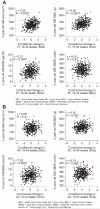
We have demonstrated previously that higher birth weight is associated with greater peak and later-life bone mineral content and that maternal body build, diet, and lifestyle influence prenatal bone mineral accrual. To examine prenatal influences on bone health further, we related ultrasound measures of fetal growth to childhood bone size and density. We derived Z-scores for fetal femur length and abdominal circumference and conditional growth velocity from 19 to 34 weeks' gestation from ultrasound measurements in participants in the Southampton Women's Survey. A total of 380 of the offspring underwent dual-energy X-ray absorptiometry (DXA) at age 4 years [whole body minus head bone area (BA), bone mineral content (BMC), areal bone mineral density (aBMD), and estimated volumetric BMD (vBMD)]. Volumetric bone mineral density was estimated using BMC adjusted for BA, height, and weight. A higher velocity of 19- to 34-week fetal femur growth was strongly associated with greater childhood skeletal size (BA: r = 0.30, p < .0001) but not with volumetric density (vBMD: r = 0.03, p = .51). Conversely, a higher velocity of 19- to 34-week fetal abdominal growth was associated with greater childhood volumetric density (vBMD: r = 0.15, p = .004) but not with skeletal size (BA: r = 0.06, p = .21). Both fetal measurements were positively associated with BMC and aBMD, indices influenced by both size and density. The velocity of fetal femur length growth from 19 to 34 weeks' gestation predicted childhood skeletal size at age 4 years, whereas the velocity of abdominal growth (a measure of liver volume and adiposity) predicted volumetric density. These results suggest a discordance between influences on skeletal size and volumetric density.
Copyright 2010 American Society for Bone and Mineral Research.
Figures
References
-
- Tanner JM. Foetus into Man: Physical Growth from Conception to Maturity. 2nd ed. Castlemead Publications; Ware, England: 1989. The organisation of the growth process; pp. 165–177.
-
- Tanner JM. Foetus into Man: Physical Growth from Conception to Maturity. 2nd ed. Castlemead Publications; Ware, England: 1989. Growth before birth; pp. 36–50.
-
- Little RE. Mother’s and father’s birthweight as predictors of infant birthweight. Paediatr Perinat Epidemiol. 1987;1:19–31. - PubMed
-
- Hanson MA, Godfrey KM. Commentary: maternal constraint is a pre-eminent regulator of fetal growth. Int J Epidemiol. 2008;37:252–254. - PubMed
-
- Hernandez CJ, Beaupre GS, Carter DR. A theoretical analysis of the relative influences of peak BMD, age-related bone loss and menopause on the development of osteoporosis. Osteoporos Int. 2003;14:843–847. - PubMed
Publication types
MeSH terms
Grants and funding
LinkOut - more resources
Full Text Sources
Medical


