

doi: 10.7861/clinmedicine.10-2-181.
Treatment of acquired valvular heart disease: percutaneous alternatives
Affiliations
- PMID: 20437997
- PMCID: PMC4952098
- DOI: 10.7861/clinmedicine.10-2-181
Item in Clipboard
Treatment of acquired valvular heart disease: percutaneous alternatives
Clin Med (Lond).
2010 Apr.
No abstract available
Figures
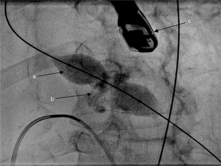
Aortic balloon valvuloplasty: (a) ‘Nucleus™’ balloon shown with ‘waisting’ at the level of the heavily calcified aortic valve (b) as it inflates. A transoesophageal echocardiography probe is seen (c), although this is not a prerequisite for aortic valvuloplasty.
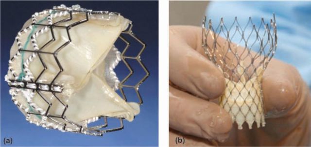
Edwards Sapien Valve (a) and CoreValve prosthesis (b) prior to loading on to the delivery system ((b) reproduced from Ref 12 with permission from Elsevier).
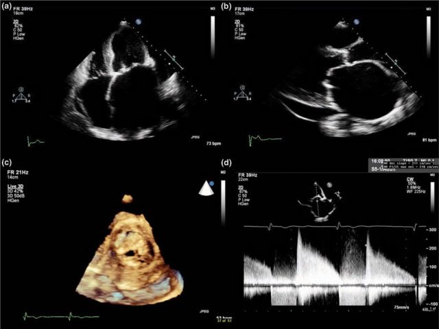
Typical echocardiographic features of mitral stenosis: limited opening of the valve with thickened and calcified leaflets, severely dilated left atrium, diastolic doming of the anterior mitral valve leaflet, decreased E-F slope of the mitral inflow Doppler tracing, increased mean pressure gradient and small valve area with planimetry: (a) apical four-chamber view; (b) parasternal long axis view; (c) mitral stenosis with 3D echocardiography; (d) mitral valve inflow Doppler tracing.
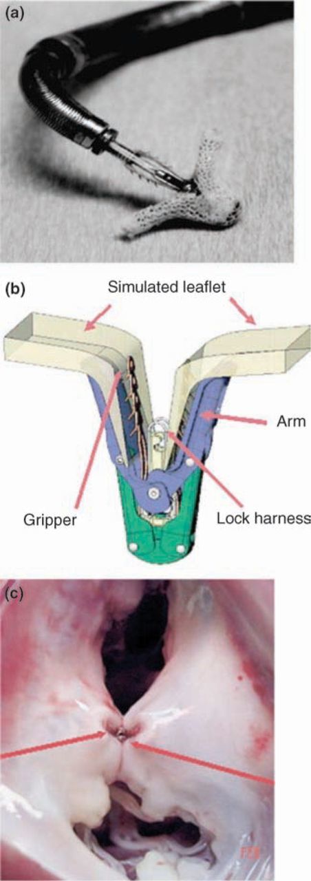
The Evalve mitral clip (Evalve, Inc): (a) clip device on distal tip of triaxial catheter delivery system; (b) illustrated components of clip device grasping mitral valve leaflet; (c) animal pathology specimen of the mitral valve with clip resulting in a double-orifice mitral valve (reproduced from Ref 17 with permission from Elsevier).
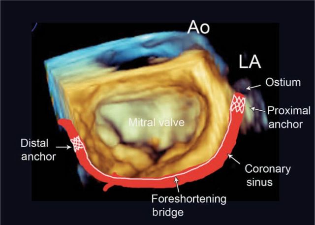
Schematic derived from a 3D echocardiographic image of the mitral valve, illustrating the location of the coronary sinus and implanted annuloplasty device. Ao = aorta; LA = left ventricle.
References
-
- Fratz S, Gildein HP, Balling G, et al. Aortic valvuloplasty in paediatric patients substantially postpones the need for aortic valve surgery: a single-centre experience of 188 patients after up to 17.5 years of follow-up. Circulation 2008;117:1201–6 - PubMed
MeSH terms
LinkOut - more resources
Full Text Sources
Medical
