Review
. 2010 Aug 15:12:203-31.
doi: 10.1146/annurev-bioeng-070909-105351.

Topography, cell response, and nerve regeneration

Affiliations
Review

Topography, cell response, and nerve regeneration

Diane Hoffman-Kim et al. Annu Rev Biomed Eng. .

Abstract

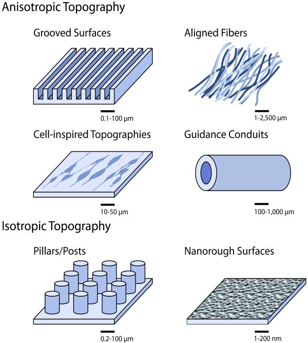
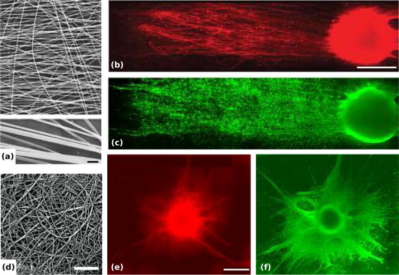
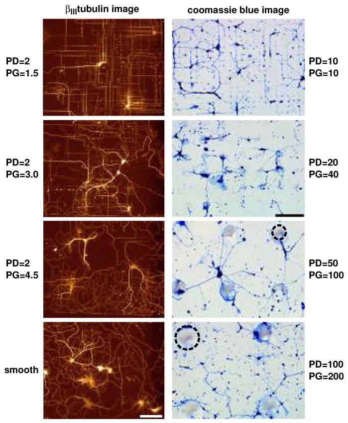
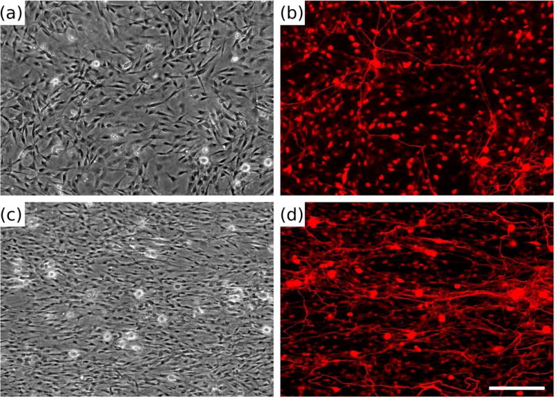
In the body, cells encounter a complex milieu of signals, including topographical cues, in the form of the physical features of their surrounding environment. Imposed topography can affect cells on surfaces by promoting adhesion, spreading, alignment, morphological changes, and changes in gene expression. Neural response to topography is complex, and it depends on the dimensions and shapes of physical features. Looking toward repair of nerve injuries, strategies are being explored to engineer guidance conduits with precise surface topographies. How neurons and other cell types sense and interpret topography remains to be fully elucidated. Studies reviewed here include those of topography on cellular organization and function as well as potential cellular mechanisms of response.


Figures

Figure 1
Figure 1
Topographies presented to neurons in vitro.
Figure 2
Figure 2
Dorsal root ganglia (DRGs) on aligned and random electrospun nanoscale fiber film in vitro. (a, d) Representative scanning electron microscopy (SEM) images of the aligned poly(acrylonitrile-comethylacrylate) fibers (a, with magnified fibers below, scale bar = 1 μm) and the random fibers (d, scale bar = 30 μm). (b, c) Double immunostained DRG on the aligned fiber film: representative montage of NF160 (a marker for axons) immunostained DRG neurons on the film (b) and montage of S-100 (a marker for Schwann cells) immunostained Schwann cells on the film (c), scale bar = 500 μm. (e, f) Double immunostained DRG on the random fiber film: representative montage of NF160+ neurons (e) and S-100+ Schwann cells (f), scale bar = 500 μm. Adapted with permission from Kim et al. (2008).
Figure 3
Figure 3
Effects of surface topography on the orientation of embryonic rat hippocampal neurons cultured on surfaces with varying pillar diameter (PD) and pillar gap (PG). Left column: Fluorescence images demonstrate the effects of poly-l-lysine-coated silicon pillars of diameter 2.0 μm at increasing pillar gap sizes. The greatest orientation was demonstrated for β III-tubulin-labeled processes on pillar gaps of 1.5 μm. Effects of orientation decreased as the gap size increased to 3.0 and 4.5 μm. Processes on smooth regions demonstrated random growth. Adapted with permission from Dowell-Mesfin et al. 2004. Right column: Optical micrographs, at the same magnification and light settings, of Coomassie blue-stained hippocampal neurons plated on glass substrates patterned with conical posts of polydimethylsiloxane (PDMS) of 10 μm diameter, 10 μm gap; 20 μm diameter, 40 μm gap; 50 μm diameter, 100 μm gap; and 100 μm diameter, 200 μm gap. Dotted circles outline representative pillars. Adapted with permission from Hanson et al. (2009).
Figure 4
Figure 4
Dorsal root ganglia (DRG) morphology on biomimetic materials presenting replicated Schwann cell (SC) topography. Phase contrast (a, c) and fluorescent micrographs (b, d) of dissociated DRG cultured for 5 days on polydimethylsiloxane (PDMS) (a, b) or replicas (c, d) and stained with neurofilament immunocytochemistry (b, d). Adapted with permission from Bruder et al. (2007).
Figure 5
Figure 5
Coordination of actin cytoskeleton on grid surface during cell spreading is inhibited by Blebbistatin and Y27632, but not ML-7. Left column: F-actin of untreated cells cultured on polydimethylsiloxane (PDMS) flat, line, or grid patterns for 26 h. Other columns: F-actin of inhibitor-treated cells spreading on patterned substrates. Inhibitors were added to cell suspension during plating and cells were fixed and stained after 2 h of incubation (red: Rhodamine-Phalloidin, blue: DAPI). Adapted with permission from Mai et al. (2007).

References

    1. Harrison RG. The cultivation of tissues in extraneous medium as a method of morphogenetic study. Anat Rec. 1912;6:181–93.
    1. Weiss P. The problem of specificity in growth and development. Yale J Bio Med. 1947;19(3):235–278. - PMC - PubMed
    1. Curtis AS, Varde M. Control of cell behavior: topological factors. J Natl Cancer Inst. 1964;33:15–26. - PubMed
    1. Voldman J, Gray ML, Schmidt MA. Microfabrication in biology and medicine. Annu Rev Biomed Eng. 1999;1:401–25. - PubMed
    1. Curtis A, Wilkinson C. Topographical control of cells. Biomaterials. 1997;18(24):1573–83. - PubMed

Publication types

Substances

LinkOut - more resources