. 2010 Aug 1;49(3):348-60.
doi: 10.1016/j.freeradbiomed.2010.04.021. Epub 2010 May 14.

Activation of ASK-1 and downstream MAP kinases in cytochrome P4502E1 potentiated tumor necrosis factor alpha liver injury

Affiliations

Activation of ASK-1 and downstream MAP kinases in cytochrome P4502E1 potentiated tumor necrosis factor alpha liver injury

Defeng Wu et al. Free Radic Biol Med. .

Abstract

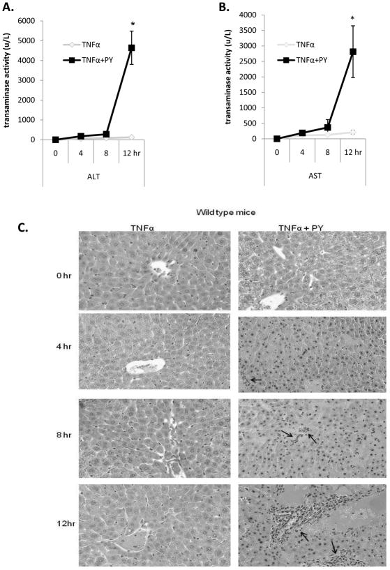
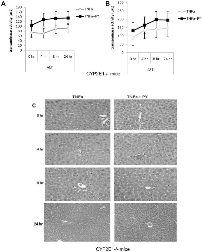
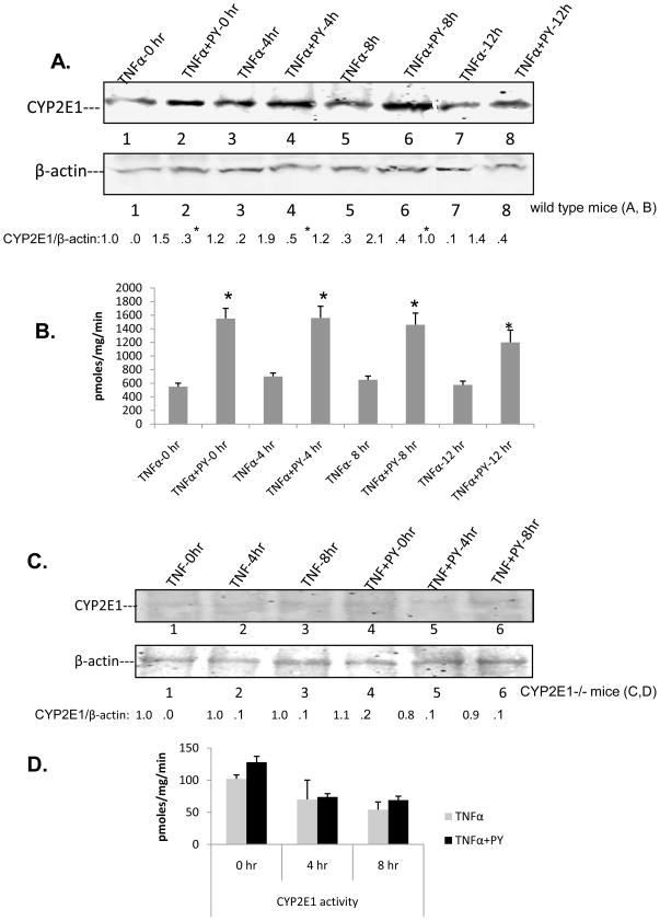
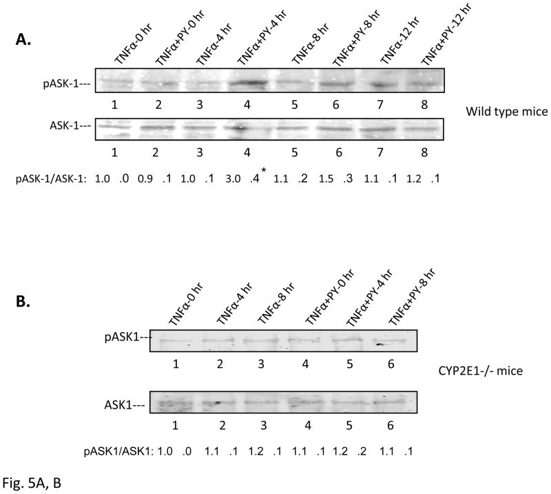
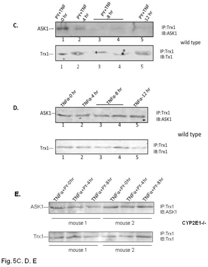
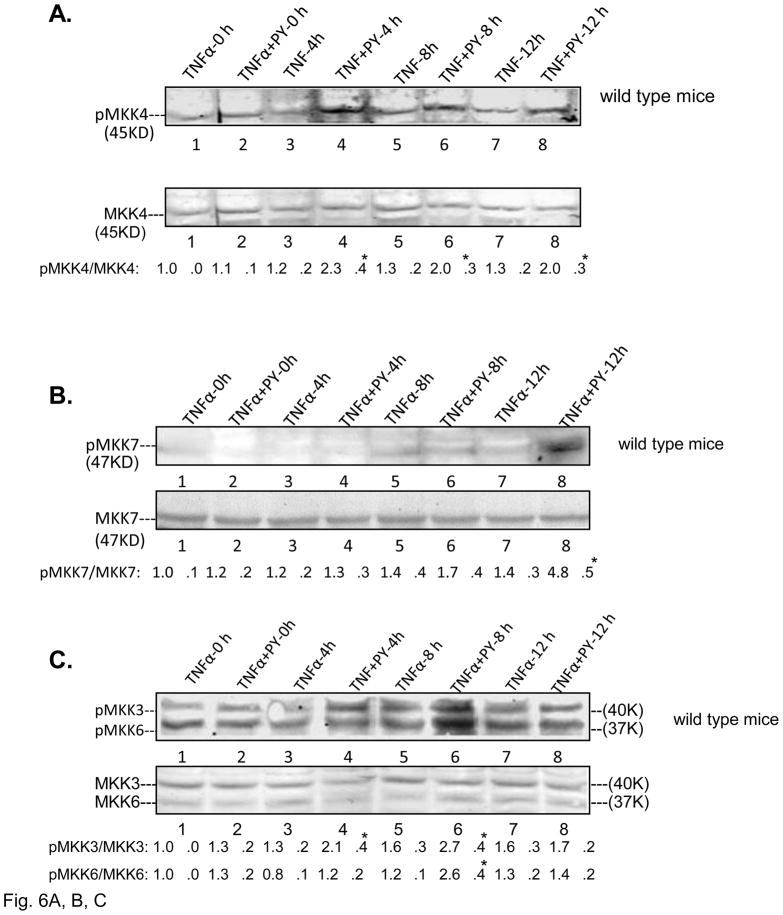
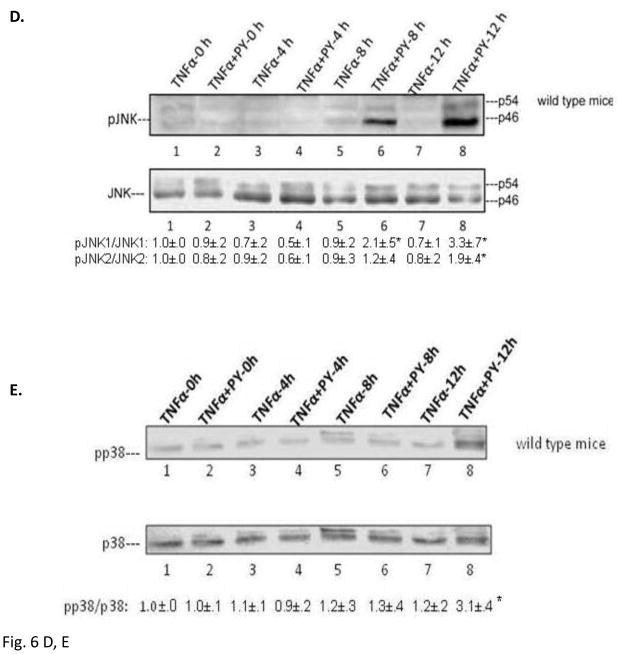
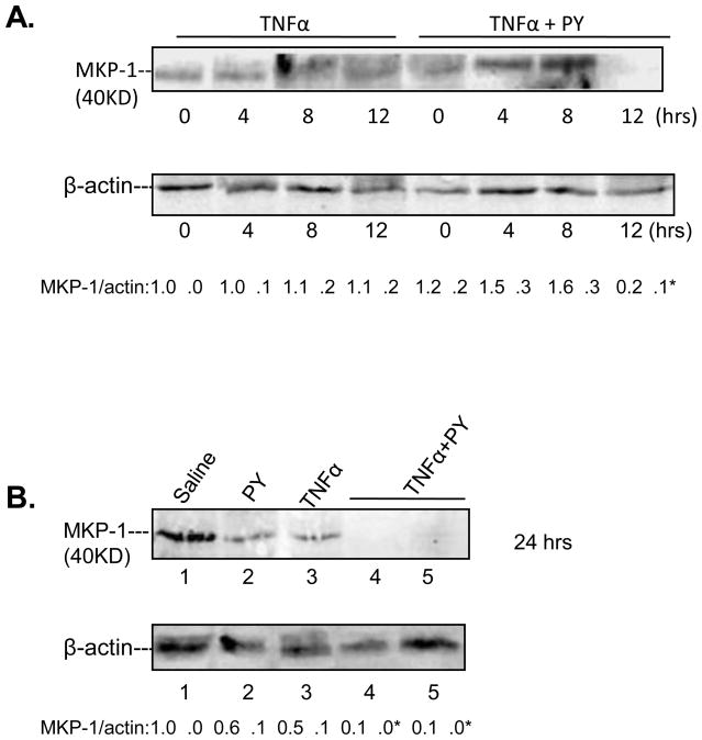
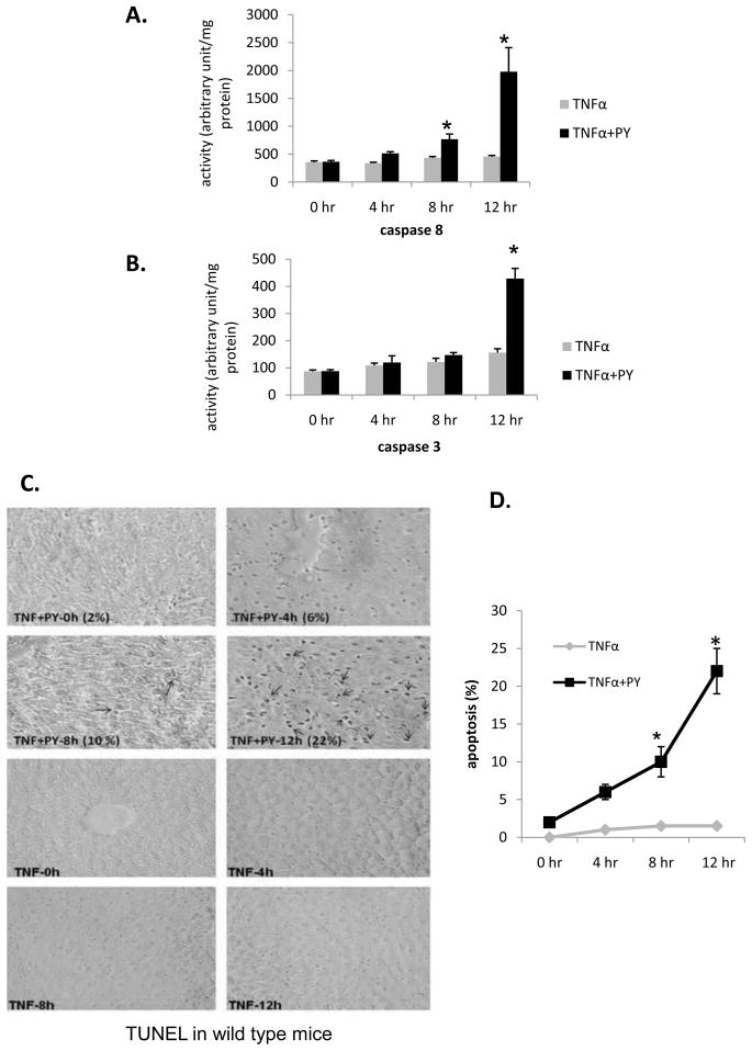
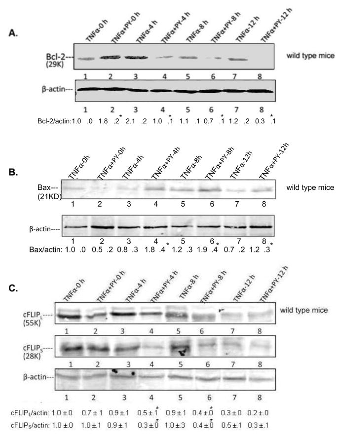
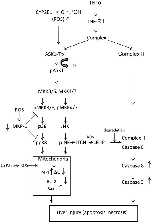
Cytochrome P4502E1 (CYP2E1) potentiates TNFalpha toxicity by a mechanism involving increased oxidative stress and activation of JNK and p38 MAPKs. This study evaluated the upstream mediators of this MAPK activation with a special focus on studying whether apoptosis signal regulating kinase-1 (ASK-1) is activated in the CYP2E1-TNFalpha hepatotoxic model. Wild-type and CYP2E1(-/-) mice were treated with pyrazole (PY) for 3days to induce CYP2E1 and challenged with TNFalpha on day 3. Liver injury occurred between 8 and 12h after TNFalpha administration only to the wild-type PY-treated mice. Oxidative stress was elevated in the PY mice at 4h, a time before the liver injury. ASK-1 was dissociated from the thioredoxin-ASK-1 complex and was activated at 4h after administration of TNFalpha to PY mice. This was followed by activation of MKK3/MKK6 and MKK4/MKK7 at 4-8 or 12h and then JNK/p38 MAPK at 8 to 12h. MAPK phosphatase-1 was decreased 12 to 24h after TNFalpha administration. This may promote a sustained activation of JNK. Bax was elevated, whereas Bcl-2 and cFLIP(S/L) were lowered at 4h after administration of TNFalpha. These changes were followed by increases in caspase 8 and 3 activities and apoptosis. None of the above changes were observed when TNFalpha was administered to PY-treated CYP2E1(-/-) mice. These studies show that TNFalpha increases oxidative stress in mice with elevated CYP2E1, with subsequent activation of ASK-1 via a mechanism involving thioredoxin-ASK-1 dissociation, followed by activation of downstream MKK and MAPK. We speculate that similar interactions between CYP2E1 and TNFalpha may be important for alcohol-induced liver injury.


Figures

Fig. 1
Fig. 1. CYP2E1 plus TNFα-dependent liver injury
Wild type mice were treated with saline or pyrazole for 3 days as described in Methods and Materials. On day 3, TNFα was administered immediately after the saline or pyrazole treatment and the mice were sacrificed at 0, 4, 8 and 12 hours, after the TNFα. ALT (A), AST (B) and histopathology (H&E staining) (C). Arrows indicate foci of inflammatory infiltration focus. Results are the average from 4 mice in each group at each time point.. P<0.05 for the 12h TNFα+PY versus TNFα.
Fig. 2
Fig. 2. Hepatotoxicity evaluation in CYP2E1−/− mice
CYP2E1−/− mice were treated as mentioned in the legend to Fig. 1 and sacrificed at 0, 4, 8 and 24h, respectively, after the TNFα. ALT (A), AST (B), H&E staining (C). Results are the average of 4 mice. There were no significant differences between the TNFα versus the TNFα plus PY.
Fig. 3
Fig. 3. CYP2E1 plus TNFα-dependent ROS stress
WT mice (A, C) or CYP2E1−/− mice (B, D) were treated as described in the legend to Fig. 1, and at the indicated times after administration of TNFα, GSH levels (A, WT; B, 2E1−/−) and lipid peroxidation (C, WT; D, 2E1−/−) evaluated. Results are each from three mice. *, P< 0.05, compared with TNFα alone at different time points.
Fig. 4
Fig. 4. CYP2E1 levels
Immunoblots for CYP2E1 and oxidation of PNP at 0, 4, 8, 12h after administration of TNFα to saline-or pyrazole-treated WT mice (A,B) or CYP2E1−/− mice (C,D). Numbers under the blots refer to the CYP2E1/β-actin ratio. For A and B, CYP2E1 levels and oxidation of PNP were significantly higher (*, P<0.05) for the TNFα+PY compared to the TNFα. Results are from 4 mice per group at each time point
Fig. 5
Fig. 5. Activation of ASK-1
The activation of ASK-1 was determined in liver lysate from WT (A) or CYP2E1−/− mice (B) by immunoblot analysis. The activation was expressed as the pASK/ASK ratio and ratios from 3 experiments are shown below the blots. * P<0.05 compared with TNFα alone at 4 H. Trx antibody was used to immunoprecipitate the Trx-ASK complex from TNFα plus PY (C) or TNFα alone (D) treated mouse liver lysates from WT (C, D) or CYP2E1−/− (E) mice. ASK-1 and Trx-1 levels in the immunoprecipitate were determined by immunoblot..
Fig. 6
Fig. 6. Activation of MKK4/MKK7, MKK3/MKK6, JNK and p38 MAPK
The activations of MKK4 (A,); MKK7 (B); MKK3, MKK6 (C); JNK (D ) and p38 MAPK (E ) in wild type mice were analyzed from liver lysates by immunoblot analysis. The activation was expressed as the phosphorylated over non-phosphorylated form and ratios from 3 different mice are shown under the blots. *, P<0.05 compared with their TNFα alone control respectively.
Fig. 7
Fig. 7. Levels of MKP-1
Immunoblots were carried out to determine MKP-1 levels in liver lysates from TNFα alone or TNFα plus PY treated wild type mice. The levels of MKP-1 were determined from the MKP-1/β-actin ratios; ratios from 3 different mice are expressed below the blots. *, P<0.05 compared with TNFα alone groups for 12h or 24h respectively.
Fig. 8
Fig. 8. Activation of caspase 8, 3 and DNA fragmentation
Caspase 8 (A) and caspase 3 (B) activation in liver lysates from wild type mice treated with saline or with pyrazole was determined at the indicated time points after administration of TNFα. * P< 0.05 compared with TNFα alone. Experiments were repeated with three different mice. Liver slices from PY plus TNFα or TNFα alone treated mice were used for TUNEL analysis as described in Methods. Arrows indicate TUNEL-positive nuclei (stained dark brown). The average percent of TUNEL-positive cells are shown in (C) and graph (D), *, P<0.05 compared with TNFα alone treated groups.
Fig. 9
Fig. 9. Levels of pro-and anti-apoptotic factors
Bcl-2 (A) and Bax (B) levels in liver lysates from wild type mice were determined by immunoblot. Blot intensities were compared with β-actin as shown below the blots. The long form and short form of cFLIP, cFLIPL and cFLIPS were also determined from liver lysates by immunoblots (C). All immunoblots were repeated with three different mice and the ratios are shown below the blots. *, P<0.05 compared with their TNFα alone control respectively.
Fig. 10
Fig. 10. Scheme. CYP2E1 sensitizes TNFα to activate ASK-1 and its downsteam signaling targets
We hypothesize that CYP2E1-generated ROS cause dissociation of ASK-1 from the Trx-ASK1 complex in the presence of TNFα. This results in activation of the MAPKKK, ASK-1, followed by activation of MAPKK and eventually JNK and p38 MAPK. The combination of elevated ROS, activated JNK and p38 MAPK, increases in proapoptotic and decrease in anti-apoptotic Bcl-2 family members promote mitochondrial dysfunction and liver injury. Apoptosis occurs from the elevation of ROS, activation of MAPK, decline in cFLIP, which contributes to the liver injury. The decrease in MKP-1, likely from CYP2E1-generated ROS, results in prolonged and sustained activation of JNK and p38 MAPK.

References

    1. Thurman GR. Mechanisms of hepatic toxicity. II Alcoholic liver injury involves activation of Kupffer cells by endotoxin. Am J Physiol. 1998;275:G605–G611. - PubMed
    1. McClain CJ, Barve S, Deaciu I, Kugelmas M, Hill D. Cytokines in alcoholic liver disease. Semin Liver Dis. 1999;19:205–219. - PubMed
    1. Yin M, Wheeler MD, Kono H, Bradford BU, Galluci RM, Luster MI, Thurman RG. Essential role of TNFα in alcohol-induced liver injury in mice. Gastroenterology. 1999;117:942–952. - PubMed
    1. Rao RK, Seth A, Sheth P. Recent Advances in Alcoholic Liver Disease I. Role of intestinal permeability and endotoxemia in alcoholic liver disease. Am J Physiol Gastrointest Liver Physiol. 2004;286:G881–884. - PubMed
    1. Thurman RG, Bradford BU, Iimuro Y, Knecht KT, Arteel GE, Yin M, Connor HD, Kadiisda M, Mason RP. The role of gut-derived bacterial toxins and free radicals in alcohol-induced liver injury. J Gastroenterol Hepatol. 1998;13:S39–S50. - PubMed

Publication types

MeSH terms