. 2010 Jul;69(7):1389-95.
doi: 10.1136/ard.2009.119776. Epub 2010 May 3.

Synovial tissue hypoxia and inflammation in vivo

Affiliations

Synovial tissue hypoxia and inflammation in vivo

C T Ng et al. Ann Rheum Dis. 2010 Jul.

Abstract

Introduction: Hypoxia is a microenvironmental feature in the inflamed joint, which promotes survival advantage for cells. The aim of this study was to examine the relationship of partial oxygen pressure in the synovial tissue (tPO(2)) in patients with inflammatory arthritis with macroscopic/microscopic inflammation and local levels of proinflammatory mediators.

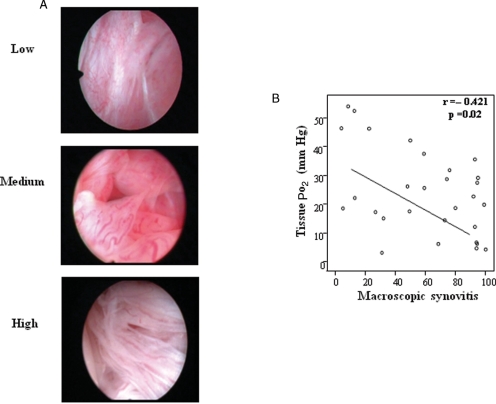
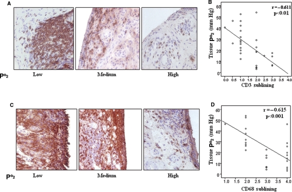
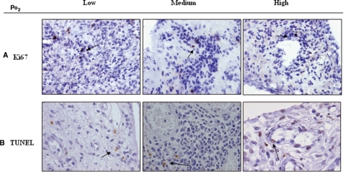
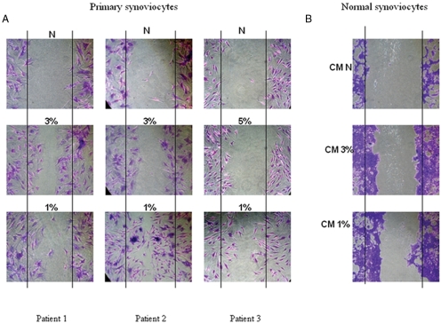
Methods: Patients with inflammatory arthritis underwent full clinical assessment and video arthroscopy to quantify macroscopic synovitis and measure synovial tPO(2) under direct visualisation. Cell specific markers (CD3 (T cells), CD68 (macrophages), Ki67 (cell proliferation) and terminal deoxynucleotidyl transferase dUTP nick end labelling (cell apoptosis)) were quantified by immunohistology. In vitro migration was assessed in primary and normal synoviocytes (synovial fibroblast cells (SFCs)) using a wound repair scratch assay. Levels of tumour necrosis factor alpha (TNFalpha), interleukin 1beta (IL1beta), interferon gamma (IFNgamma), IL6, macrophage inflammatory protein 3alpha (MIP3alpha) and IL8 were quantified, in matched serum and synovial fluid, by multiplex cytokine assay and ELISA.

Results: The tPO(2) was 22.5 (range 3.2-54.1) mm Hg and correlated inversely with macroscopic synovitis (r=-0.421, p=0.02), sublining CD3 cells (-0.611, p<0.01) and sublining CD68 cells (r=-0.615, p<0.001). No relationship with cell proliferation or apoptosis was found. Primary and normal SFCs exposed to 1% and 3% oxygen (reflecting the median tPO(2) in vivo) induced cell migration. This was coupled with significantly higher levels of synovial fluid tumour necrosis factor alpha (TNFalpha), IL1beta, IFNgamma and MIP3alpha in patients with tPO(2) <20 mm Hg (all p values <0.05).

Conclusions: This is the first study to show a direct in vivo correlation between synovial tPO(2), inflammation and cell migration, thus it is proposed that hypoxia is a possible primary driver of inflammatory processes in the arthritic joint.


Conflict of interest statement

Competing Interests: None.

Figures

Figure 1
Figure 1
A. Representative images of macroscopic synovitis in patients with low, moderate and high measures of in vivo tissue PO2 (tPO2) levels. B. Scattergraph demonstrating the inverse correlation between tPO2 and macroscopic synovitis. r, Spearman's correlation coefficient, p<0.05 is statistically significant.
Figure 2
Figure 2
Microscopic synovitis and tissue PO2 (tPO2). A,C. Representative images of immunohistochemical staining for CD3 and CD68, in lining and sublining layers, from patients with low, moderate and high in vivo tPO2 levels. B,D. The correlation between tPO2 and CD3 and CD68 staining in synovial sublining layers. r, Spearman's correlation coefficient, p<0.05 is statistically significant.
Figure 3
Figure 3
Ki67 and terminal deoxynucleotidyl transferase dUTP nick end labelling (TUNEL) staining in synovial tissues. A,B. Representative images of immunohistological staining for Ki67 (top panel) and TUNEL (lower panel), in lining and sublining layers, in patients with low, moderate and high in vivo tissue PO2 (tPO2) levels, demonstrating low expression indices of both markers.
Figure 4
Figure 4
Migration assays in primary and normal synoviocytes. A. Representative images of primary synoviocytes wound repair assays exposed to normoxia (n=3), in vivo oxygen levels (n=2 at 3%, n=1 at 5%), and positive control of 1% oxygen for 24 h. Significant migration was observed when primary synoviocytes (patients 1 and 2) were exposed to 3% when compared to normoxia (A, left and middle panels), an effect that was greater at 1% hypoxia. When primary synovial fibroblast cells (SFCs) were exposed to 5% hypoxia (patient 3) migration was minimal and similar to normoxia (A right panel), migration was demonstrated when exposed to 1%. B. Normal synoviocytes cultured in conditioned supernatant from primary SFCs exposed to normoxia, 3% and 1% oxygen for 24 h before scratches. Significant migration was observed in those incubated with conditioned media from 1% and 3% hypoxic conditions. CM, cultured media; N, normoxia.
Figure 5
Figure 5
Proinflammatory cytokines/chemokines in synovial fluid and tissue PO2 (tPO2). A–D. Significant differences in tumour necrosis factor (TNF)α, interleukin (IL)1β, interferon (IFN)γ and macrophage inflammatory protein (MIP)3α levels between patients with tPO2 values of greater or less than 20 mm Hg. p<0.05 was significantly different. E,F. No significant differences in IL6 and IL8 levels between patients with tPO2 values of greater or less than 20 mm Hg.

Similar articles

Cited by

References

    1. Alarcón GS. Epidemiology of rheumatoid arthritis. Rheum Dis Clin North Am 1995;21:589–604 - PubMed
    1. O'Neill T, Silman AJ. Psoriatic arthritis. Historical background and epidemiology. Baillieres Clin Rheumatol 1994;8:245–61 - PubMed
    1. Harris ED Jr Rheumatoid arthritis. Pathophysiology and implications for therapy. N Engl J Med 1990;322:1277–89 - PubMed
    1. Månsson B, Carey D, Alini M, et al. Cartilage and bone metabolism in rheumatoid arthritis. Differences between rapid and slow progression of disease identified by serum markers of cartilage metabolism. J Clin Invest 1995;95:1071–7 - PMC - PubMed
    1. Kruithof E, Baeten D, De Rycke L, et al. Synovial histopathology of psoriatic arthritis, both oligo- and polyarticular, resembles spondyloarthropathy more than it does rheumatoid arthritis. Arthritis Res Ther 2005;7:R569–80 - PMC - PubMed

Publication types

MeSH terms