. 2010 Jul 2;285(27):21175-84.
doi: 10.1074/jbc.M110.112482. Epub 2010 May 3.

Smooth and cardiac muscle-selective knock-out of Kruppel-like factor 4 causes postnatal death and growth retardation

Affiliations

Smooth and cardiac muscle-selective knock-out of Kruppel-like factor 4 causes postnatal death and growth retardation

Tadashi Yoshida et al. J Biol Chem. .

Abstract

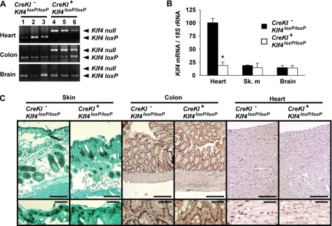
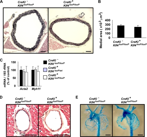
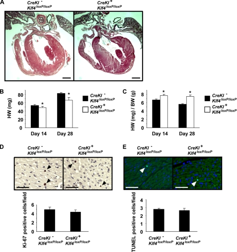
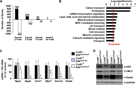
Krüppel-like factor 4 (Klf4) is a transcription factor involved in differentiation and proliferation in multiple tissues. We demonstrated previously that tamoxifen-induced deletion of the Klf4 gene in mice accelerated neointimal formation but delayed down-regulation of smooth muscle cell differentiation markers in carotid arteries following injury. To further determine the role of Klf4 in the cardiovascular system, we herein derived mice deficient for the Klf4 gene in smooth and cardiac muscle using the SM22alpha promoter (SM22alpha-CreKI(+)/Klf4(loxP/loxP) mice). SM22alpha-CreKI(+)/Klf4(loxP/loxP) mice were born at the expected Mendelian ratio, but they gradually died after birth. Although approximately 40% of SM22alpha-CreKI(+)/Klf4(loxP/loxP) mice survived beyond postnatal day 28, they exhibited marked growth retardation. In wild-type mice, Klf4 was expressed in the heart from late embryonic development through adulthood, whereas it was not expressed in smooth muscle. No changes were observed in morphology or expression of smooth muscle cell differentiation markers in vessels of SM22alpha-CreKI(+)/Klf4(loxP/loxP) mice. Of interest, cardiac output was significantly decreased in SM22alpha-CreKI(+)/Klf4(loxP/loxP) mice, as determined by magnetic resonance imaging. Moreover, a lack of Klf4 in the heart resulted in the reduction in expression of multiple cardiac genes, including Gata4. In vivo chromatin immunoprecipitation assays on the heart revealed that Klf4 bound to the promoter region of the Gata4 gene. Results provide novel evidence that Klf4 plays a key role in late fetal and/or postnatal cardiac development.


Figures

FIGURE 1.
FIGURE 1.
Smooth and cardiac muscle-specific deletion of the Klf4 gene was associated with significant postnatal death and growth retardation. A, a schematic representation of smooth and cardiac muscle-specific deletion of the Klf4 gene is shown. The numbers shown represent Klf4 exons. Triangles represent the loxP sites. X, breeding. B, the genotype of 191 offspring from the breeding between SM22α-CreKI+ (CreKI)/Klf4loxP/wt mice and SM22α-CreKI/Klf4loxP/loxP mice was examined by PCR at the time of birth. C, Kaplan-Meier survival curves for SM22α-CreKI+/Klf4loxP/loxP, SM22α-CreKI+/Klf4loxP/wt, SM22α-CreKI/Klf4loxP/loxP, and SM22α-CreKI/Klf4loxP/wt mice are shown (n = 36 per each genotype). A log-rank test for trend yielded. *, p < 0.05. D and E, representative pictures of SM22α-CreKI+/Klf4loxP/loxP and SM22α-CreKI/Klf4loxP/loxP mice at P1 (D) and P28 (E) are shown. F, changes in the body weight of SM22α-CreKI+/Klf4loxP/loxP, SM22α-CreKI+/Klf4loxP/wt, SM22α-CreKI/Klf4loxP/loxP, and SM22α-CreKI/Klf4loxP/wt mice after birth are shown (n = 20∼25 per each genotype). *, p < 0.05 compared with other genotypes. Docta represent the mean ± S.E.
FIGURE 2.
FIGURE 2.
Klf4 was expressed in the heart from late embryonic development to adulthood. A–F, Klf4 expression was determined by immunohistochemistry in wild-type embryos at E9.5 (A), at E11.5 (B), at E15.5 (C), and at E18.5 (D), as well as in the heart of wild-type neonates at P1 (E) and adult (F). Klf4 expression was visualized by DAB, and sections were counterstained with hematoxylin. Bars for A–C, 100 μm; D–F, 50 μm. H, heart; DA, dorsal aorta; III, third branchial arch artery; IV, fourth branchial arch artery; VI, sixth branchial arch artery. G, colocalization of Klf4 and Gata4 in the heart of wild-type mice was examined by dual immunofluorescence staining. Klf4 staining was visualized by Alexa Fluor 555 (red), and Gata4 staining was visualized by Alexa Fluor 488 (green). Sections were counterstained with 4′,6-diamidino-2-phenylindole (blue). Arrowheads indicate cells coexpressing Klf4 and Gata4. Bar, 50 μm. H–K, Klf4 expression was examined in the aorta (H), the skeletal muscle (I), the stomach (J), and the bladder (K) of wild-type mice. Arrowheads indicate aortic endothelial cells (H) and gastric epithelial cells (J), which express Klf4. SM, smooth muscle layer. Bars for H and I, 50 μm; bars for J and K, 100 μm. Representative pictures are shown.
FIGURE 3.
FIGURE 3.
SM22α-CreKI+/Klf4loxP/loxP mice exhibited selective loss of Klf4 expression in the heart. A, deletion of the Klf4loxP allele was tested in multiple tissues including the heart, the colon, and the brain of SM22α-CreKI/Klf4loxP/loxP and SM22α-CreKI+/Klf4loxP/loxP mice at P28, as determined by PCR (n = 3 per each genotype). B, expression of Klf4 mRNA was examined by real-time RT-PCR in the heart, the skeletal muscle (sk. m), and the brain of SM22α-CreKI/Klf4loxP/loxP and SM22α-CreKI+/Klf4loxP/loxP mice at P28 (n = 6 per each genotype). C, Klf4 expression was examined by immunohistochemistry in the skin, the colon, and the heart of SM22α-CreKI/Klf4loxP/loxP and SM22α-CreKI+/Klf4loxP/loxP mice at P28. Klf4 was visualized by DAB, and sections were counterstained with azure B for the skin or with hematoxylin for the colon and the heart. Representative pictures are shown (n = 4 per each genotype). Bars, 100 μm for upper panels; 50 μm for lower panels.
FIGURE 4.
FIGURE 4.
SM22α-CreKI+/Klf4loxP/loxP mice had no differences in expression of SMC differentiation marker genes or arterial structure as compared with control mice. A and B, the thoracic aorta from SM22α-CreKI/Klf4loxP/loxP and SM22α-CreKI+/Klf4loxP/loxP mice at P28 was perfused with 10% formalin under physiological pressure and embedded into paraffin. Cross-sections were stained with Russell-Movat pentachrome method (n = 6 per each genotype). A, representative pictures are shown. Bars, 100 μm. B, medial areas (the average of three sections per each mouse) were measured using ImagePro software. C, expression of SMC differentiation marker genes including Acta2 and Myh11 was examined by real-time RT-PCR in the aorta of SM22α-CreKI/Klf4loxP/loxP, SM22α-CreKI+/Klf4loxP/wt, and SM22α-CreKI+/Klf4loxP/loxP mice at P14 (n = 5 per each genotype). D, coronary arteries from SM22α-CreKI/Klf4loxP/loxP and SM22α-CreKI+/Klf4loxP/loxP mice at P28 was sectioned and stained with Russell-Movat pentachrome method (n = 6 per each genotype). Representative pictures of the left anterior descending artery are shown. Bars, 50 μm. E, vascular casting of coronary arteries from E18.5 embryos is shown. Bars, 1 mm.
FIGURE 5.
FIGURE 5.
The rates of proliferation and apoptosis were unaltered in the hearts of SM22α-CreKI+/Klf4loxP/loxP mice. A, hearts from SM22α-CreKI/Klf4loxP/loxP and SM22α-CreKI+/Klf4loxP/loxP mice at P28 were sectioned and stained with Russell-Movat pentachrome method. Representative pictures are shown (n = 6 per each genotype). Bars, 1 mm. B and C, heart weight and the ratio of heart to body weight were measured in SM22α-CreKI/Klf4loxP/loxP and SM22α-CreKI+/Klf4loxP/loxP mice at P14 and P28. *, p < 0.05 compared with SM22α-CreKI/Klf4loxP/loxP mice. D and E, Ki-67 staining (D) and TUNEL staining (E) were performed in the heart of SM22α-CreKI/Klf4loxP/loxP and SM22α-CreKI+/Klf4loxP/loxP mice at P28 (n = 5 per each genotype). Ki-67 staining was visualized by DAB, and sections were counterstained with hematoxylin. TUNEL staining was visualized by fluorescein isothiocyanate, and sections were counterstained with 4′,6-diamidino-2-phenylindole. Arrowheads indicate stained cells. Bars, 50 μm.
FIGURE 6.
FIGURE 6.
Multiple cardiac genes were decreased in the heart of SM22α-CreKI+/Klf4loxP/loxP mice. A and B, microarray analysis was performed in the heart of SM22α-CreKI/Klf4loxP/loxP and SM22α-CreKI+/Klf4loxP/loxP mice at P14 (n = 4 per each genotype). A, number of genes dysregulated by Klf4 deletion is shown by fold change. B, the 427 genes increased or decreased >3-fold by Klf4 deletion were subjected to gene ontology analysis with PANTHER. Significantly enriched biological processes (p < 0.03) are shown. Plotted is the log (p value) with the threshold set to 1.5 [-log(0.03)]. C, expression of multiple cardiac genes including Nppa, Nppb, Actc1, Myh7, Serca2, and Gata4 was examined by real-time RT-PCR in the heart of SM22α-CreKI/Klf4loxP/loxP, SM22α-CreKI+/Klf4loxP/wt, and SM22α-CreKI+/Klf4loxP/loxP mice at P14 (n = 5 per each genotype). D, protein expression of Klf4, cardiac myosin light chain (cMLC), Gata4, and glyceraldehyde-3-phosphate dehydrogenase (GAPDH) was examined in the heart of SM22α-CreKI/Klf4loxP/loxP and SM22α-CreKI+/Klf4loxP/loxP mice at P14 by Western blotting. Representative pictures are shown (n = 3 for each genotype).
FIGURE 7.
FIGURE 7.
Klf4 regulated Gata4 expression by binding to the Gata4 promoter region. A, a schematic diagram of the Gata4 promoter is shown. A consensus Klf4 binding site, 5′-(G/A)(G/A)GG(C/T)G(C/T)-3′, is located within the Gata4 promoter, which contains 4 GC-rich elements and an E box. Numbers represent bp. B, association of Klf4 with the promoter regions of the Gata4 gene and the c-fos gene was determined by in vivo ChIP assays in the heart of SM22α-CreKI/Klf4loxP/loxP and SM22α-CreKI+/Klf4loxP/loxP mice at P14 (n = 3). *, p < 0.05 compared with control mice. Ab, antibody.

Similar articles

Cited by

References

    1. Segre J. A., Bauer C., Fuchs E. (1999) Nat. Genet. 22, 356–360 - PubMed
    1. Katz J. P., Perreault N., Goldstein B. G., Lee C. S., Labosky P. A., Yang V. W., Kaestner K. H. (2002) Development 129, 2619–2628 - PMC - PubMed
    1. Katz J. P., Perreault N., Goldstein B. G., Actman L., McNally S. R., Silberg D. G., Furth E. E., Kaestner K. H. (2005) Gastroenterology 128, 935–945 - PubMed
    1. Swamynathan S. K., Katz J. P., Kaestner K. H., Ashery-Padan R., Crawford M. A., Piatigorsky J. (2007) Mol. Cell. Biol. 27, 182–194 - PMC - PubMed
    1. Takahashi K., Tanabe K., Ohnuki M., Narita M., Ichisaka T., Tomoda K., Yamanaka S. (2007) Cell 131, 861–872 - PubMed

Publication types

MeSH terms

LinkOut - more resources