. 2010 Jul 2;285(27):20904-14.
doi: 10.1074/jbc.M109.098558. Epub 2010 May 3.

Phosphorylation of mixed lineage leukemia 5 by CDC2 affects its cellular distribution and is required for mitotic entry

Affiliations

Phosphorylation of mixed lineage leukemia 5 by CDC2 affects its cellular distribution and is required for mitotic entry

Jie Liu et al. J Biol Chem. .

Abstract

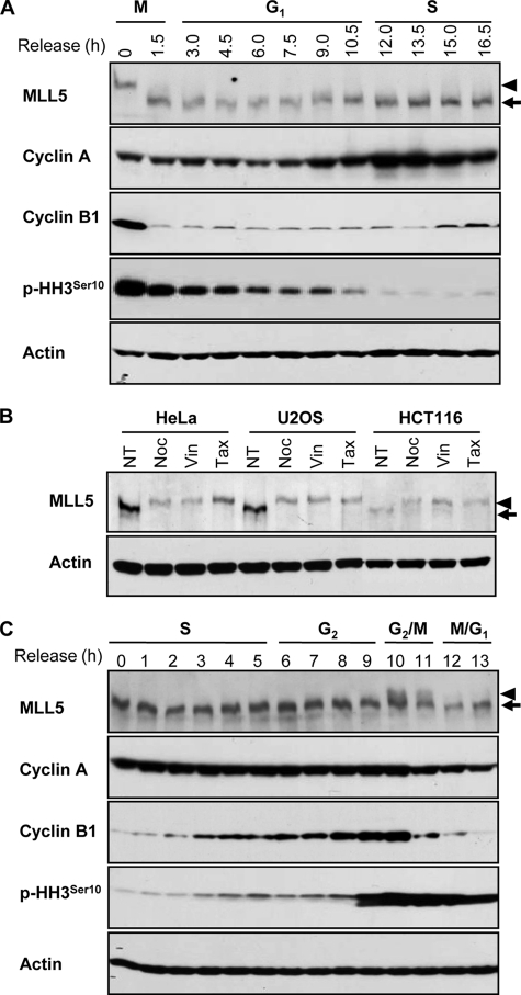
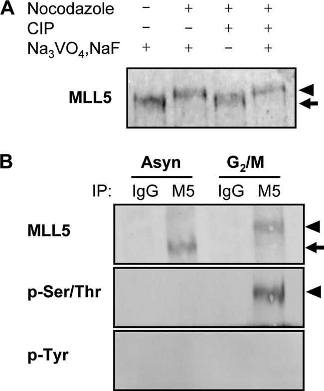
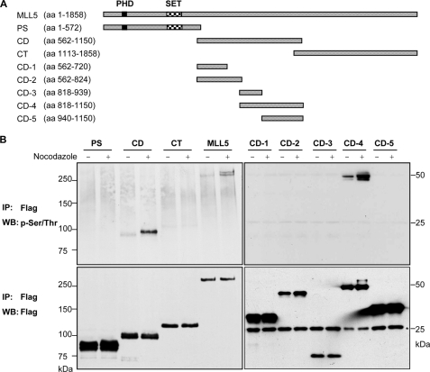
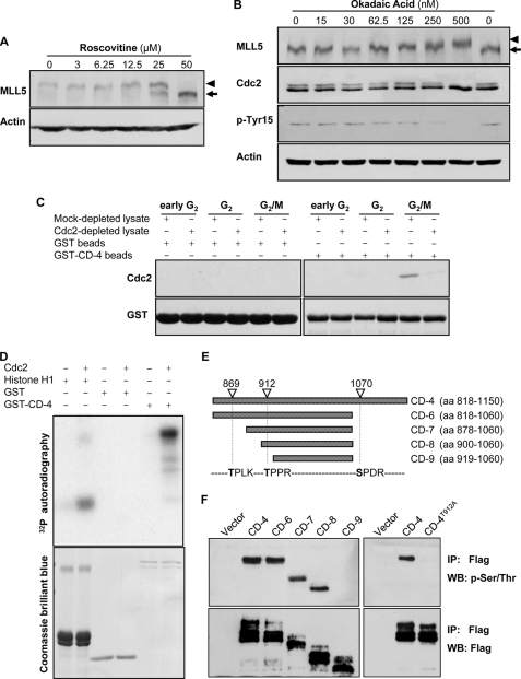
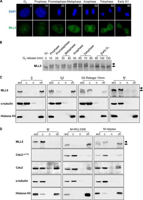
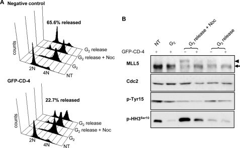
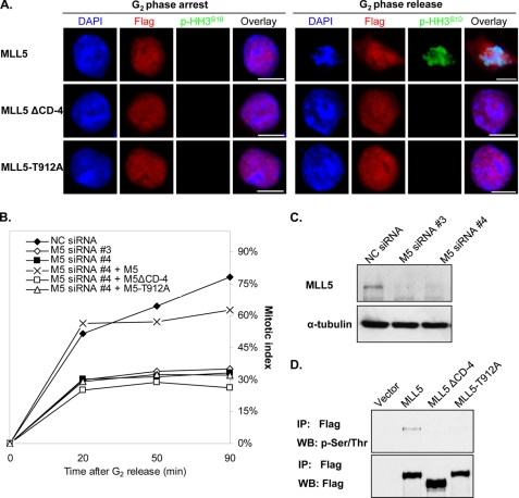
The human mixed lineage leukemia-5 (MLL5) gene is frequently deleted in myeloid malignancies. Emerging evidence suggests that MLL5 has important functions in adult hematopoiesis and the chromatin regulatory network, and it participates in regulating the cell cycle machinery. Here, we demonstrate that MLL5 is tightly regulated through phosphorylation on its central domain at the G(2)/M phase of the cell cycle. Upon entry into mitosis, the phosphorylated MLL5 delocalizes from condensed chromosomes, whereas after mitotic exit, MLL5 becomes dephosphorylated and re-associates with the relaxed chromatin. We further identify that the mitotic phosphorylation and subcellular localization of MLL5 are dependent on Cdc2 kinase activity, and Thr-912 is the Cdc2-targeting site. Overexpression of the Cdc2-targeting MLL5 fragment obstructs mitotic entry by competitive inhibition of the phosphorylation of endogenous MLL5. In addition, G(2) phase arrest caused by depletion of endogenous MLL5 can be compensated by exogenously overexpressed full-length MLL5 but not the phosphodomain deletion or MLL5-T912A mutant. Our data provide evidence that MLL5 is a novel cellular target of Cdc2, and the phosphorylation of MLL5 may have an indispensable role in the mitotic progression.


Figures

FIGURE 1.
FIGURE 1.
MLL5 displays slower gel mobility at G2/M transition. A, expression of MLL5 in HeLa cells released from the G2/M boundary. B, expression of MLL5 in asynchronous (no treatment (NT)) and G2/M-arrested HeLa, U2OS, and HCT116 cells. G2/M synchronization was achieved by treatment with nocodazole (Noc), vinblastine (Vin), or taxol (Tax). C, expression of MLL5 in HeLa cells that were released from G1/S synchronization. Cyclin A, cyclin B1, and phosphohistone H3Ser10 serve as cell cycle progression markers. Actin serves as a protein loading control. Arrows indicate MLL5, and arrowheads denote the slower migrating form of MLL5 at G2/M phase.
FIGURE 2.
FIGURE 2.
MLL5 is phosphorylated at Ser/Thr residues at G2/M phase. A, alkaline phosphatase treatment of G2/M-arrested HeLa cell extract converted MLL5 to a faster migrating form that ran to the same migrating position as in the asynchronous cell extract. CIP, calf intestinal alkaline phosphatase. B, endogenous MLL5 was immunoprecipitated (IP) from asynchronous or G2/M-arrested HeLa cell extracts. Phosphorylated MLL5 (arrowheads) is present at G2/M phase and can be detected by anti-phospho-Ser/Thr but not anti-phospho-Tyr antibodies.
FIGURE 3.
FIGURE 3.
CD-4 domain of MLL5 is sufficient to mediate mitotic phosphorylation. A, schematic representation of MLL5 and deletion fragments; PS, PHDSET domain; CD, central domain; CT, C-terminal domain. The central domain was further dissected into CD-1, CD-2, CD-3, CD-4, and CD-5. aa, amino acids. B, full-length MLL5 and various deletion mutants were immunoprecipitated (IP) from untreated or nocodazole-synchronized HEK 293T cell lysates with anti-FLAG antibodies and detected by anti-phospho-Ser/Thr or anti-FLAG antibodies. MLL5 was phosphorylated at Ser/Thr residues on the CD-4 domain at G2/M phase. The numbers indicate the molecular masses (kDa) of the protein standards, and the asterisk denotes the light chain of antibodies. WB, Western blot.
FIGURE 4.
FIGURE 4.
MLL5 is phosphorylated by Cdc2 at G2/M. A, mitotic HeLa cells were treated with various concentrations of roscovitine for 4 h in the presence of nocodazole. The phosphorylation of MLL5 was inhibited by roscovitine in a dose-dependent manner. B, asynchronous HeLa cells were treated with various concentrations of okadaic acid for 4 h. The phosphorylation of MLL5 was gradually induced by okadaic acid in a dose-dependent manner. C, GST-CD-4 protein co-purified with Cdc2 in HeLa cells that were released from the G1/S phase boundary for 12 h (G2/M). D, upper panel, GST-CD-4 was phosphorylated by human recombinant Cdc2-cyclin B1 complex in an in vitro kinase assay. Histone H1 and GST served as a positive and a negative control, respectively. Lower panel, Coomassie Brilliant Blue staining for histone H1, GST, and GST-CD-4. E, schematic representation of MLL5-CD-4 domain and its deletion fragments. The potential Cdc2 targeting sites on MLL5-CD-4 domain are denoted based on the consensus motif for Cdc2 kinase ((S/T)PX(K/R)). aa, amino acids. F, left panel, CD-4 domain and deletion mutants (CD-6–9) with sequential deletion of Cdc2-targeting sites were immunoprecipitated (IP) from nocodazole-synchronized extract of HEK 293T cells using anti-FLAG antibodies and detected by anti-phospho-Ser/Thr or anti-FLAG antibodies. The phosphorylation on MLL5 is abrogated when Thr-912 residue was deleted. Right panel, mutation of Thr-912 to Ala on the CD-4 domain eliminated the phosphorylation modification, suggesting that Thr-912 is the phosphorylation targeting site by Cdc2. WB, Western blot.
FIGURE 5.
FIGURE 5.
Phospho-MLL5 dissociates from mitotic chromosomes. HeLa cells were grown on coverslips, and G2-arrested cells were released into fresh medium. Samples for immunofluorescence staining and Western blotting were collected every 10 min. A, MLL5 formed intranuclear foci in G2 phase (time 0), but it dissociated from condensed chromosomes and displayed cytosolic staining pattern during mitosis (10 min, prophase; 20 min, prometaphase; 30 min, metaphase; 50 min, anaphase; and 60 min, telophase). When cells completed mitosis (90 min), MLL5 re-localized to the nucleus. Scale bar, 10 μm. DAPI, 4′,6-diamidino-2-phenylindole. B, time course study on the phosphorylation of MLL5 during mitosis. C, HeLa cells were arrested at difference cell cycle stages, and cellular fractionation was performed. Cells arrested in S phase were collected after G1/S release for 4 h; G2 phase arrest was achieved by incubation with RO-3306 for 20 h, and M phase cells were synchronized by the Thy-nocodazole method. α-Tubulin and histone H3 were employed as cytoplasmic (c) and chromatin-associated (ch) protein marker, respectively, and nucleoplasmic protein was denoted as group (n). Whole cell lysate (wcl) serves as total cellular protein control. Phosphorylation and subcellular localization of MLL5 were examined by Western blotting. MLL5 was extracted in the chromatin fraction in S and G2 phase cells (1st and 2nd panel), and in mitotic cells the MLL5 was phosphorylated and extracted in the cytoplasmic fraction (3rd and 4th panels). D, mitotic phosphorylation and localization of MLL5 were dependent on Cdc2 kinase activity. In mitosis-arrested HeLa cells, MLL5 was phosphorylated and extracted in the cytoplasmic fraction (left panel). When mitotic HeLa cells were treated with RO-3306 for 1.5 h, MLL5 was dephosphorylated and extracted in the chromatin fraction. Inhibition of Cdc2 activity was revealed by the increase in phosphorylation of Cdc2 on Tyr-15 (middle panel). When mitotic cells were released into complete medium for 1.5 h and re-entered into the G1 phase, MLL5 was dephosphorylated and associated with chromatin (right panel). Phospho-MLL5 was denoted by an arrowhead.
FIGURE 6.
FIGURE 6.
Inhibition of endogenous MLL5 phosphorylation impedes mitotic progression and arrests cells at the G2/M transition. HEK 293T cells were transfected with GFP-CD-4 in the absence (no treatment (NT)) or presence of RO-3306 for 20 h before release into complete medium or nocodazole-containing medium for 5 h. A, cell cycle analysis for GFP-negative control cells and GFP-CD-4-expressing cells. The percentage represents the population that successfully progressed through mitosis and re-entered G1 phase after 5 h release from G2 arrest. B, population of GFP-CD-4 positive or GFP-negative cells released from RO-3306 was sorted separately before harvesting for Western blotting. Phosphorylation of endogenous MLL5 at G2/M phase (G2 release + nocodazole (Noc)) was inhibited by ectopic overexpression of GFP-CD-4. Arrowhead indicates phospho-MLL5; arrow indicates MLL5.
FIGURE 7.
FIGURE 7.
G2 arrest caused by the depletion of endogenous MLL5 can be rescued by exogenous expression of FLAG-MLL5 but not FLAG-MLL5ΔCD-4 or FLAG-MLL5-T912A. Endogenous MLL5 expression in HEK 293T cells were knocked down by siRNA, and FLAG-MLL5, FLAG-MLL5ΔCD-4, or FLAG-MLL5-T912A was transfected to rescue the cell cycle arrest. Cells were synchronized to G2 phase and allowed for mitotic progression. MLL5 localization was analyzed by immunofluorescence staining using anti-FLAG antibodies. Mitotic index was calculated by counting the phosphohistone H3Ser10-positive cells at 20, 50, and 90 min post-release. A, FLAG-MLL5 protein dissociated from chromosomes and the cells entered mitosis after G2 release for 20 min, as shown by positive histone H3Ser10 staining and chromosome condensation (1st row). FLAG-MLL5ΔCD-4 and FLAG-MLL5-T912A were restricted in nuclei, and there was no visible chromatin condensation, which was marked by histone H3Ser10 phosphorylation. Scale bar, 10 μm. DAPI, 4′,6-diamidino-2-phenylindole. B, cumulative mitotic index was calculated for control cells (NC siRNA), endogenous MLL5-knockdown cells (M5 siRNA #3 or M5 siRNA #4), FLAG-MLL5-positive cells (M5 siRNA #4 + M5), FLAG-MLL5ΔCD-4-positive cells (M5 siRNA #4 + M5ΔCD-4), and FLAG-MLL5-T912A-positive cells (M5 siRNA #4 + M5-T912A). C, Western blotting showed the successful knockdown of endogenous MLL5 by siRNA numbers 3 and 4. D, immunoprecipitation result showed that FLAG-MLL5ΔCD-4 and FLAG-MLL5-T912A could not be phosphosphorylated upon nocodazole treatment.

Similar articles

Cited by

References

    1. Hasle H., Aricò M., Basso G., Biondi A., Cantù Rajnoldi A., Creutzig U., Fenu S., Fonatsch C., Haas O. A., Harbott J., Kardos G., Kerndrup G., Mann G., Niemeyer C. M., Ptoszkova H., Ritter J., Slater R., Starý J., Stollmann-Gibbels B., Testi A. M., van Wering E. R., Zimmermann M. (1999) Leukemia 13, 376–385 - PubMed
    1. Brezinová J., Zemanová Z., Ransdorfová S., Pavlistová L., Babická L., Housková L., Melichercíková J., Sisková M., Cermák J., Michalová K. (2007) Cancer Genet. Cytogenet. 173, 10–16 - PubMed
    1. Luna-Fineman S., Shannon K. M., Lange B. J. (1995) Blood 85, 1985–1999 - PubMed
    1. Johnson E. J., Scherer S. W., Osborne L., Tsui L. C., Oscier D., Mould S., Cotter F. E. (1996) Blood 87, 3579–3586 - PubMed
    1. Le Beau M. M., Espinosa R., 3rd, Davis E. M., Eisenbart J. D., Larson R. A., Green E. D. (1996) Blood 88, 1930–1935 - PubMed

Publication types

MeSH terms