. 2010 Jul 2;285(27):20532-40.
doi: 10.1074/jbc.M110.109298. Epub 2010 May 3.

MicroRNA-340-mediated degradation of microphthalmia-associated transcription factor mRNA is inhibited by the coding region determinant-binding protein

Affiliations

MicroRNA-340-mediated degradation of microphthalmia-associated transcription factor mRNA is inhibited by the coding region determinant-binding protein

Srikanta Goswami et al. J Biol Chem. .

Retraction in

Abstract

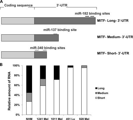
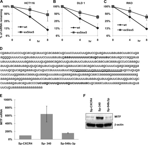
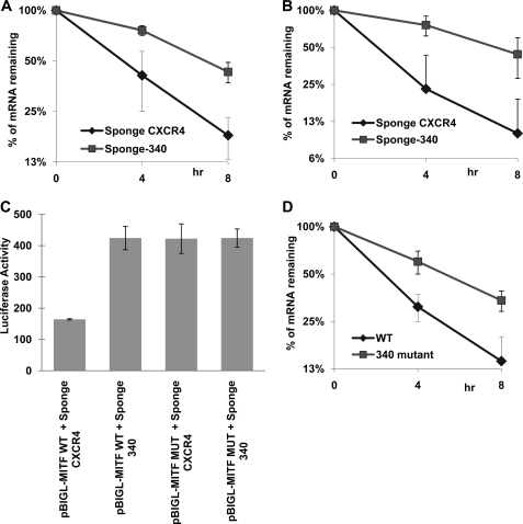
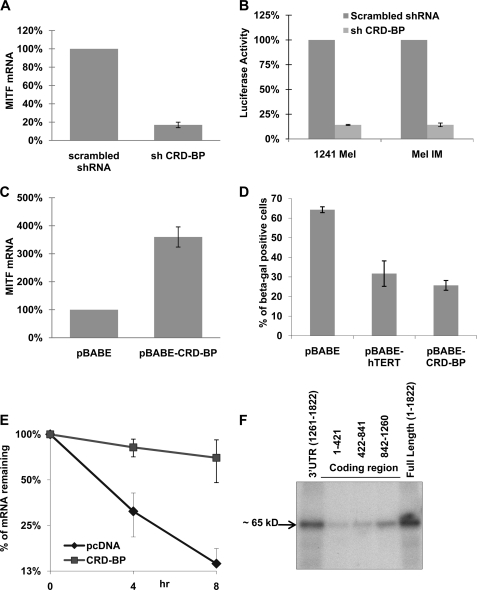
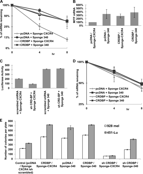
Alternative cleavage and polyadenylation generate multiple transcript variants of mRNA isoforms with different length of 3'-untranslated region (UTR). Alternative cleavage and polyadenylation enable differential post-transcriptional regulation of transcripts via the availability of different cis-acting elements in 3'-UTRs. Microphthalmia-associated transcription factor (MITF) is a master regulator of melanocyte development and melanogenesis. It has also been implicated in melanoma development. Here we show that melanoma cells favor the expression of MITF mRNA with shorter 3'-UTR. This isoform of mRNA is regulated by microRNA, miR-340. miR-340 interacts with two of its target sites on the 3'-UTR of MITF mRNA, causing mRNA degradation and decreased expression and activity of MITF. On the other hand, the RNA-binding protein coding region determinant-binding protein, shown to be highly expressed in melanoma, directly binds to the 3'-UTR of MITF mRNA and prevents the binding of miR-340 to its target sites, resulting in stabilization of the MITF transcript and elevated expression and transcriptional activity of MITF. This interplay between RNA-binding protein and miRNA describes the important mechanism of regulation of MITF in melanocytes and malignant melanomas.


Figures

FIGURE 1.
FIGURE 1.
MITF mRNA with short 3′-UTR is more abundant in melanoma cell lines. A, graphical representation of different MITF mRNA isoforms with varying lengths of 3′-UTRs. The approximate positions of the reported miRNAs are shown on long and medium 3′-UTRs. B, real time quantitative PCR with primers specific for long, medium, and short 3′-UTRs (see “Experimental Procedures” for primer sequence) using RNA extracted from NHMs and the indicated melanoma cell lines.
FIGURE 2.
FIGURE 2.
The abundant MITF mRNA with short 3′-UTR is also regulated by miRNA. A, Dicerwt and DicerEx5/Ex5 HCT116 cells were co-transfected with Tet-off plasmid and p-BIG-MITF plasmid with short 3′-UTR. Transcription was stopped by treatment with doxycycline for the indicated durations. The stability of MITF transcripts was analyzed by measuring MITF mRNA levels with real time RT-PCR (normalized to GAPDH expression). B, p-BIG-MITF plasmid with short 3′-UTR was expressed in DLD1 cells, Dicerwt (wt), and DicerEx5/Ex5 (ex5/ex5) under the control of the Tet-off system. The stability of MITF mRNA was analyzed as in A. C, stability of MITF transcript with short 3′-UTR expressed in RKOwt and RKO DicerEx5/Ex5 cells was analyzed as in A. All of the results are representative of three separate experiments and are expressed as the mean values ± S.D. (error bars). The average half-lives of mRNAs are presented in supplemental Table S3. D, sequence of the short 3′-UTR of MITF showing binding sites for miR-340 (in bold type) and miR-548c-3p (underlined). E, the levels of endogenous MITF mRNA in 451Lu cells, transfected with the indicated miR-Sponge constructs, were estimated by real time RT-PCR after normalization with respect to GAPDH expression. The results are representative of three separate experiments and are expressed as percentages of control (SP-CXCR4) as the mean values ± S.D. (error bars). F, immunoblot analysis of MITF expression in the 451Lu cells transfected with the indicated miR-Sponge constructs (upper panel). The lower panel shows the expression of β-actin. The levels of endogenous miR-340 are presented in supplemental Fig. S1A.
FIGURE 3.
FIGURE 3.
MITF mRNA is a target of miR-340. A, 451Lu cells were co-transfected with Tet-off plasmid, p-BIG-MITF plasmid with short 3′-UTR, and the indicated miR-Sponge construct. Transcription was stopped by treatment with doxycycline for the indicated durations. The stability of MITF transcripts was analyzed by measuring MITF mRNA levels with real time RT-PCR (normalized to GAPDH expression). B, 451Lu cells were co-transfected with Tet-off plasmid, p-BIGL-MITF-Short MITF 3′-UTR, and the indicated miR-Sponge construct. The turnover of chimeric Luciferase-MITF-3′-UTR transcripts was analyzed as in A. C, 451Lu cells were co-transfected with β-galactosidase-expressing plasmid, Tet-off plasmid, p-BIGL-MITFwt, or p-BIGL-MITFδmiR-340 and the indicated miR-Sponge constructs. After 24 h the luciferase activity was measured. The values represent luciferase activity normalized to β-galactosidase. D, 451Lu cells were co-transfected with Tet-off plasmid, p-BIG-MITFwt, or p-BIG-MITFδmiR-340. The stability of MITF transcripts was analyzed as in A. All of the results are representative of three separate experiments and are expressed as the mean values ± S.D. (error bars). The average half-lives of mRNAs are presented in supplemental Table S3. The levels of endogenous miR-340 are presented in supplemental Fig. S1A.
FIGURE 4.
FIGURE 4.
CRD-BP is a positive regulator of MITF expression. A, 451Lu cells were transfected with either control shRNA or shRNA against CRD-BP. 48 h after transfection, the cells were collected and assayed for levels of MITF mRNA by quantitative RT-PCR, normalized to GAPDH expression, and presented as percentages of control (scrambled shRNA). B, 1241 Mel and Mel IM cells were co-transfected with the indicated shRNA-expressing plasmids, β-galactosidase-expressing plasmid, and pHTRPL4, where the luciferase gene is expressed under the MITF-dependent promoter. The values corresponding to luciferase activity normalized to β-galactosidase expression are presented. C, NHMs were electroporated using AMAXATM with either CRD-BP-overexpressing plasmid or empty vector. 72 h after transfection, the cells were collected, assayed for levels of MITF mRNA by quantitative RT-PCR normalized to GAPDH expression, and presented as percentages of control (pBABE). D, NHM cells were electroporated using AMAXATM with indicated plasmids. 48 h after electroporation, the cells were stained for senescence-associated β-galactosidase, and the percentages of β-galactosidase-positive cells were calculated. E, 451Lu cells were co-transfected with Tet-off plasmid, p-BIG-MITF, and either pcDNA control or CRD-BP-expressing plasmid. The turnover of MITF transcript was analyzed by real time RT-PCR after stopping transcription by doxycycline treatment for the indicated time points. Normalization was done with respect to GAPDH expression. All of the results are representative of three separate experiments and are expressed as the mean values ± S.D. (error bars). The average half-lives of mRNAs are presented in supplemental Table S3. F, FLAG immunoprecipitation of UV cross-linked ribonuclear protein complexes. Protein extracts from 293T cells, transfected with FLAG-CRDBP, were incubated with internally 32P-labeled RNA transcripts of three fragments of the MITF mRNA coding region, MITF full-length mRNA, and the short 3′-UTR. Fragment 1 contains nucleotides 1–421 of the coding region, fragment 2 consists of nucleotides 422–841, and fragment 3 consists of nucleotides 842–1260 of the MITF coding region. Ribonuclear protein complexes were precipitated with anti-FLAG antibodies, analyzed on PAGE, and autoradiographed.
FIGURE 5.
FIGURE 5.
CRD-BP inhibition reduces mitfa expression and pigmentation in zebrafish embryos. A, zebrafish embryos were injected with CRD-BP morpholino (5 or 10 ng) or standard control morpholino. Expression levels of the zebrafish MITF ortholog, mitfa, were assayed by real time quantitative PCR at 24 h post-fertilization and normalized to zebrafish α-tubulin. Morphant expression levels are presented relative to levels of expression in control embryos. B, representative zebrafish embryo at 48 h post-fertilization, injected with control or CRD-BP morpholino. CRD-BP morphants have less pigmentation and reduced numbers of pigment cells (melanophores) in 33 of 33 morphants (from seven independent experiments).
FIGURE 6.
FIGURE 6.
CRD-BP counteracts miR-340 action and stabilizes MITF mRNA. A, wild type MITF-expressing plasmid p-BIG-MITFwt was co-transfected with Tet-off plasmid and the indicated constructs in 451Lu cells. The turnover of MITF transcript was analyzed as Fig. 4E. B, 451Lu cells were transfected with the indicated constructs. 48 h after transfection, the cells were collected and assayed for levels of endogeneous MITF mRNA by quantitative RT-PCR, normalized to GAPDH expression, and presented as percentages of control. C, 451Lu cells were co-transfected with MITF-dependent luciferase-expressing vector pHTRPL4, the indicated shRNA and miR-Sponge-expressing plasmids, and β-galactosidase-expressing plasmid. The values correspond to luciferase activity normalized to β-galactosidase expression. D, construct expressing MITF with both of the miR-340 sites deleted (p-BIG-MITFδmiR-340) was co-transfected with Tet-off plasmid and the indicated constructs in 451Lu cells. The turnover of MITF transcript was analyzed as Fig. 4E. All of the results are representative of three separate experiments and are expressed as the mean values ± S.D. (error bars). The average half-lives of mRNAs are presented in supplemental Table S3. E, 451Lu and 928 Mel cells were co-transfected with indicated constructs and pTk-Puro. The colonies were selected for puromycin resistance, stained with crystal violet, and counted.

References

    1. Hallsson J. H., Haflidadóttir B. S., Stivers C., Odenwald W., Arnheiter H., Pignoni F., Steingrímsson E. (2004) Genetics 167, 233–241 - PMC - PubMed
    1. Bentley N. J., Eisen T., Goding C. R. (1994) Mol. Cell Biol. 14, 7996–8006 - PMC - PubMed
    1. Hemesath T. J., Steingrímsson E., McGill G., Hansen M. J., Vaught J., Hodgkinson C. A., Arnheiter H., Copeland N. G., Jenkins N. A., Fisher D. E. (1994) Genes Dev. 8, 2770–2780 - PubMed
    1. Yasumoto K., Yokoyama K., Shibata K., Tomita Y., Shibahara S. (1994) Mol. Cell Biol. 14, 8058–8070 - PMC - PubMed
    1. Carreira S., Liu B., Goding C. R. (2000) J. Biol. Chem. 275, 21920–21927 - PubMed

Publication types

MeSH terms

LinkOut - more resources