. 2010 Jul;30(13):3233-48.
doi: 10.1128/MCB.01178-09. Epub 2010 May 3.

Extracellular signal-regulated kinase promotes Rho-dependent focal adhesion formation by suppressing p190A RhoGAP

Affiliations

Extracellular signal-regulated kinase promotes Rho-dependent focal adhesion formation by suppressing p190A RhoGAP

Ashok K Pullikuth et al. Mol Cell Biol. 2010 Jul.

Abstract

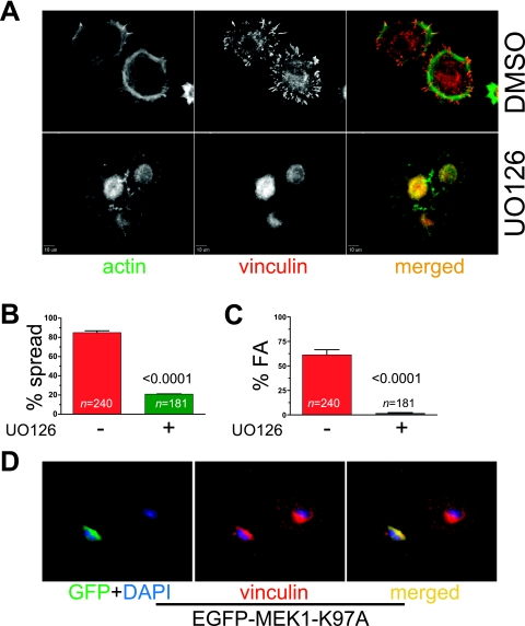
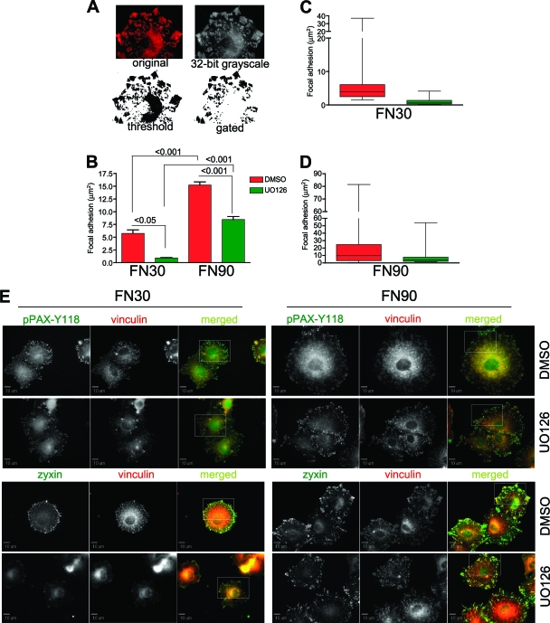
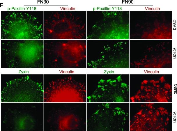
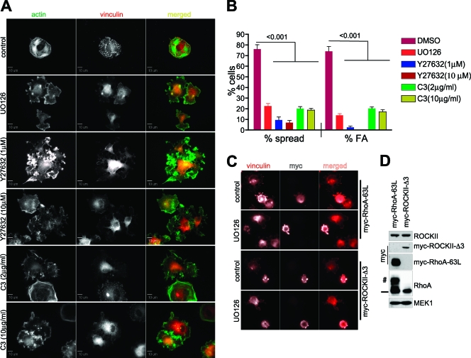
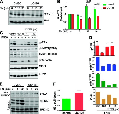
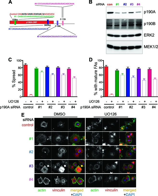
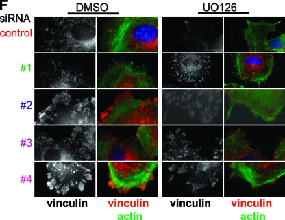
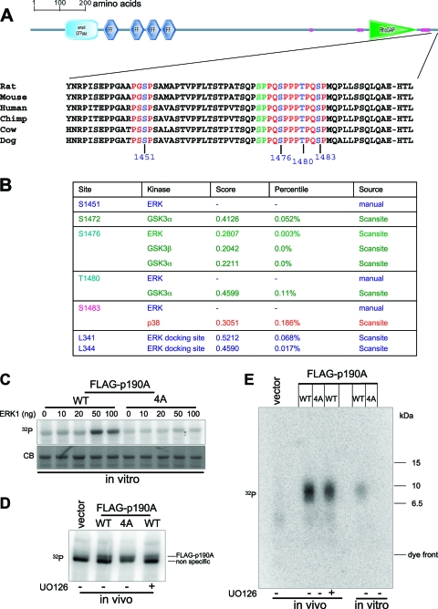
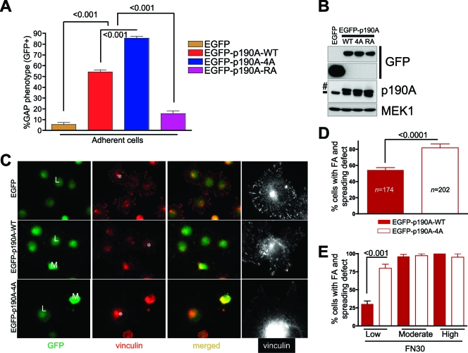
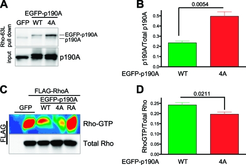
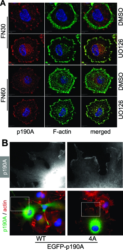
Cell migration is critical for normal development and for pathological processes including cancer cell metastasis. Dynamic remodeling of focal adhesions and the actin cytoskeleton are crucial determinants of cell motility. The Rho family and the mitogen-activated protein kinase (MAPK) module consisting of MEK-extracellular signal-regulated kinase (ERK) are important regulators of these processes, but mechanisms for the integration of these signals during spreading and motility are incompletely understood. Here we show that ERK activity is required for fibronectin-stimulated Rho-GTP loading, Rho-kinase function, and the maturation of focal adhesions in spreading cells. We identify p190A RhoGAP as a major target for ERK signaling in adhesion assembly and identify roles for ERK phosphorylation of the C terminus in p190A localization and activity. These observations reveal a novel role for ERK signaling in adhesion assembly in addition to its established role in adhesion disassembly.


Figures

FIG. 1.
FIG. 1.
MEK activity is required for cell spreading and focal adhesion assembly. REF52 cells were plated on fibronectin-coated coverslips for 30 min with or without prior treatment with the MEK inhibitor UO126 (25 μM). Cells were fixed and stained for focal adhesions (vinculin) and F-actin (phalloidin-Oregon green 488) (A) and scored for spreading (B) and for mature focal adhesions (C). At least 20 random fields were counted for each experimental condition. Experiments were performed on at least three different occasions with similar results. Data were analyzed by Student's t test, and P values and number of cells (n) in this experiment are indicated. (D) REF52 cells were transfected with EGFP-tagged kinase-defective MEK1 (EGFP-MEK1-K97A) and replated on fibronectin-coated coverslips for 30 min. Cells were fixed and imaged as in panel A.
FIG. 2.
FIG. 2.
MEK signaling facilitates maturation of focal adhesions. Cells plated on fibronectin for 30 or 90 min (FN30 and FN90, respectively) in the presence or absence of UO126 were stained for vinculin. Digital images were converted to 32-bit gray scale, threshold set and semiautomatically gated (A). Pixel values were converted to micrometers and analyzed by one-way ANOVA (B). (C and D) Box-whisker plot representation of the distribution of focal adhesion areas in cells plated for 30 of 90 min, respectively. The top and bottom edges of the rectangle are 75th and 25th percentiles, and the horizontal line within the rectangle is the median value. The means ± standard errors (SE) from this analysis are presented in panel B. (E) Cells allowed to spread on fibronectin for 30 or 90 min (FN30 and FN90, respectively) in the presence or absence of UO126 were stained for zyxin, a marker for mature adhesions, or phospho-Y118 paxillin, found in both mature and nascent adhesions. (F) Cropped and magnified details from insets shown in panel E.
FIG. 2.
FIG. 2.
MEK signaling facilitates maturation of focal adhesions. Cells plated on fibronectin for 30 or 90 min (FN30 and FN90, respectively) in the presence or absence of UO126 were stained for vinculin. Digital images were converted to 32-bit gray scale, threshold set and semiautomatically gated (A). Pixel values were converted to micrometers and analyzed by one-way ANOVA (B). (C and D) Box-whisker plot representation of the distribution of focal adhesion areas in cells plated for 30 of 90 min, respectively. The top and bottom edges of the rectangle are 75th and 25th percentiles, and the horizontal line within the rectangle is the median value. The means ± standard errors (SE) from this analysis are presented in panel B. (E) Cells allowed to spread on fibronectin for 30 or 90 min (FN30 and FN90, respectively) in the presence or absence of UO126 were stained for zyxin, a marker for mature adhesions, or phospho-Y118 paxillin, found in both mature and nascent adhesions. (F) Cropped and magnified details from insets shown in panel E.
FIG. 3.
FIG. 3.
MEK inhibition phenocopies Rho or ROCK inhibition during acute spreading. Cells in suspension were treated with MEK inhibitor UO126, ROCK inhibitor Y27632, or the Rho inhibitor C3-transferase at the indicated concentration and then allowed to adhere and spread on fibronectin for 60 min. Cells were stained for vinculin and F-actin (A). The fractions of cells spreading normally and exhibiting mature focal adhesions were quantitated (B). All images were at the same magnification, and at least 20 random fields were counted for each experimental condition. (C) The requirement for MEK activity is upstream of Rho and ROCK activities. REF52 cells were transfected with constitutively active RhoA (myc-RhoA-63L) or ROCK (myc-ROCKII-Δ3) and replated on fibronectin-coated coverslips (30 min) with or without prior treatment with UO126. Fixed cells were stained for myc epitope (to identify transfected cells) and costained with vinculin. (D) Western blot of expression of endogenous and exogenous proteins from the experiment shown in panel C. The symbols # and — denote exogenous and endogenous RhoA, respectively.
FIG. 4.
FIG. 4.
MEK activity is required for RhoGTP loading and ROCK activity during adhesion to fibronectin. (A) Suspended REF52 cells were treated with dimethyl sulfoxide (DMSO) or UO126 and replated on fibronectin. RhoGTP was pulled down with the Rho binding domain of Rhotekin. (B) Quantitation of RhoGTP normalized to total Rho from experiments represented in panel A (n = 3). (C) Cells replated on fibronectin for 30 min (FN30) with or without prior treatment with UO126, Y27632, or DMSO vehicle were assessed for ROCK-dependent phosphorylation of MYPT (pMYPT1-T696 or -T853) and cofilin (pS3-Cofilin). (D) Quantitation of experiments represented in panel C. Values from FN30 were normalized to those in control suspended cells for each condition. (E and F) Inhibition of MEK activity increases p190A RhoGAP binding to active RhoA. Cells were treated with DMSO or UO126 and replated on fibronectin. Lysates were subjected to GST-RhoA-63L pulldown and blotted with p190A antibody to determine binding of endogenous p190A-RhoGAP to active RhoA. (F) Quantitation of p190A bound to RhoGTP at 20 min expressed as fold increase over suspended cells (0 min).
FIG. 5.
FIG. 5.
p190A RhoGAP is a major target of MEK signaling during focal adhesion maturation and cell spreading. Cells transfected with stealth siRNA targeting one of four different regions in the p190A open reading frame or 3′UTR, or a mismatched nontargeting control (see panel A; mismatched residues in the control are indicated in black letters) were treated with DMSO or UO126 and replated on fibronectin-coated coverslips for 30 min. (B) Stealth p190A siRNAs suppress p190A expression without altering expression of the closely related p190B homologue. (C and D) Quantitation of cell spreading and focal adhesions. Cells in at least 20 nonoverlapping fields were counted and subjected to one-way ANOVA. (E and F) Cells were imaged for focal adhesions or F-actin. Selected regions from images in panel E are magnified in panel F. Numbers 1 to 4 denote the siRNA used (see panel A).
FIG. 5.
FIG. 5.
p190A RhoGAP is a major target of MEK signaling during focal adhesion maturation and cell spreading. Cells transfected with stealth siRNA targeting one of four different regions in the p190A open reading frame or 3′UTR, or a mismatched nontargeting control (see panel A; mismatched residues in the control are indicated in black letters) were treated with DMSO or UO126 and replated on fibronectin-coated coverslips for 30 min. (B) Stealth p190A siRNAs suppress p190A expression without altering expression of the closely related p190B homologue. (C and D) Quantitation of cell spreading and focal adhesions. Cells in at least 20 nonoverlapping fields were counted and subjected to one-way ANOVA. (E and F) Cells were imaged for focal adhesions or F-actin. Selected regions from images in panel E are magnified in panel F. Numbers 1 to 4 denote the siRNA used (see panel A).
FIG. 6.
FIG. 6.
p190A RhoGAP is an ERK substrate. (A) Four potential ERK phosphorylation motifs are highly conserved in the p190A C terminus. Vertebrate p190A sequences are aligned to show potential ERK phosphorylation (PXSP and PXTP) motifs. (B) Collation of putative phosphorylation and docking sites identified either manually or through computational methods (ScanSite). (C and D) p190A is phosphorylated on one or more putative C-terminal ERK sites in vitro and in vivo. FLAG-tagged wild-type (WT) or p190A-4A mutant (4A; where the four putative ERK sites shown in panel A have been replaced with alanines) was immunoprecipitated, pretreated with alkaline phosphatase, and then phosphorylated in vitro with recombinant active ERK in the presence of [γ-32P]ATP (C). Alternatively, REF52 cells transfected with FLAG-p190A, FLAG-p190A-4A, or empty vector were metabolically labeled with 32Pi and FLAG-p190A forms were recovered by immunoprecipitation (D). (E) Tryptic digests of in vitro- and in vivo-labeled p190A were resolved by electrophoresis (with low-molecular-mass markers) and transferred to membrane. The positions of the dye front and ∼6.5-, 10-, and 15-kDa markers are indicated. 32P denotes phosphorimages, and CB in panel C represents a Coomassie blue-stained image of the same blot.
FIG. 7.
FIG. 7.
Mutation of C-terminal ERK phosphorylation sites increases p190A RhoGAP biological activity. REF52 cells were transfected with EGFP vector, EGFP-p190A, EGFP-p190A-4A, or catalytically inactive EGFP-p190A-RA. Twenty-four hours after transfection, cells were scored for the GAP phenotype (see text). (B) Expression of endogenous (—) and exogenous (#) proteins in cells used in panel A. (C to E) Cells transfected with EGFP-p190A constructs were replated on fibronectin-coated coverslips for 30 min, fixed, and imaged for GFP and vinculin. Expression levels of EGFP-p190A were optically determined; low and moderate expressors contained about 2 and 5 times more GFP fluorescence than background, respectively. Gray scale images are magnified images of cells indicated by asterisks. (D to E) Quantitation of focal adhesion (FA) and spreading defects. Data from the entire GFP-positive populations are shown in panel D; further analysis reveals that the frequency of focal adhesion and spreading defects is greatest in a subpopulation expressing low levels of p190A-4A (E).
FIG. 8.
FIG. 8.
Mutation of C-terminal ERK phosphorylation sites increases p190A biochemical activity in adherent cells. (A) Mutation of putative C-terminal ERK sites stimulates p190A RhoGTP binding activity. Cells were transfected with either EGFP vector, EGFP-p190A (WT) or EGFP-p190A-4A. Binding of wild-type and mutant p190A RhoGAP to RhoGTP was assessed by pulldown with GST-RhoA63L. (B) Ratio of active p190A in GST-RhoA-63L pulldown to total p190A in the starting extract. Digital images from 10 experiments were statistically analyzed. (C) Mutation of putative C-terminal ERK sites modestly stimulates p190A GAP activity in cells. Cells were cotransfected with FLAG-RhoA and EGFP, EGFP-p190A, EGFP-p190-4A, or EGFP-p190A-RA (GAP-deficient R1258A mutant). FLAG-RhoGTP levels were determined by GST-Rhotekin Rho binding domain pulldown assay. Rhotekin pulldown was blotted with FLAG antibody and pseudocolored for presentation. (D) Digital images from five experiments were statistically analyzed.
FIG. 9.
FIG. 9.
ERK signaling regulates localization of p190A in spreading cells. (A) REF52 cells were treated with UO126 or vehicle control and replated on fibronectin for 30 or 60 min (FN30 and FN60, respectively). Fixed cells were stained for endogenous p190A and F-actin. (B) COS1 cells transfected with EGFP-p190A or EGFP-p190A-4A were replated on fibronectin for 60 min, fixed, and imaged for F-actin (red) and GFP (green) fluorescence. The gray scale images correspond to GFP fluorescence magnified from the areas indicated by white boxes in the color images.

Similar articles

Cited by

References

    1. Aktories, K., and I. Just. 1995. In vitro ADP-ribosylation of Rho by bacterial ADP-ribosyltransferases. Methods Enzymol. 256:184-195. - PubMed
    1. Amano, M., K. Chihara, K. Kimura, Y. Fukata, N. Nakamura, Y. Matsuura, and K. Kaibuchi. 1997. Formation of actin stress fibers and focal adhesions enhanced by Rho-kinase. Science 275:1308-1311. - PubMed
    1. Amano, M., H. Mukai, Y. Ono, K. Chihara, T. Matsui, Y. Hamajima, K. Okawa, A. Iwamatsu, and K. Kaibuchi. 1996. Identification of a putative target for Rho as the serine-threonine kinase protein kinase N. Science 271:648-650. - PubMed
    1. Arthur, W. T., and K. Burridge. 2001. RhoA inactivation by p190RhoGAP regulates cell spreading and migration by promoting membrane protrusion and polarity. Mol. Biol. Cell 12:2711-2720. - PMC - PubMed
    1. Arthur, W. T., L. A. Petch, and K. Burridge. 2000. Integrin engagement suppresses RhoA activity via a c-Src-dependent mechanism. Curr. Biol. 10:719-722. - PubMed

Publication types

MeSH terms

LinkOut - more resources