. 2010 Jul;30(13):3216-32.
doi: 10.1128/MCB.01008-09. Epub 2010 May 3.

Histone H2B C-terminal helix mediates trans-histone H3K4 methylation independent of H2B ubiquitination

Affiliations

Histone H2B C-terminal helix mediates trans-histone H3K4 methylation independent of H2B ubiquitination

Mahesh B Chandrasekharan et al. Mol Cell Biol. 2010 Jul.

Abstract

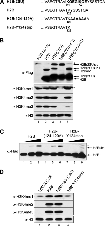
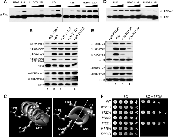
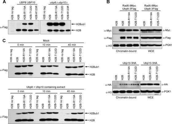
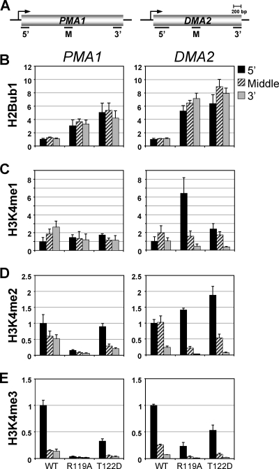
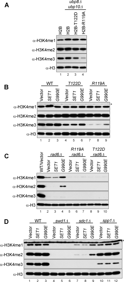
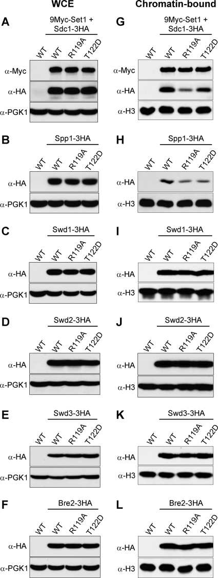
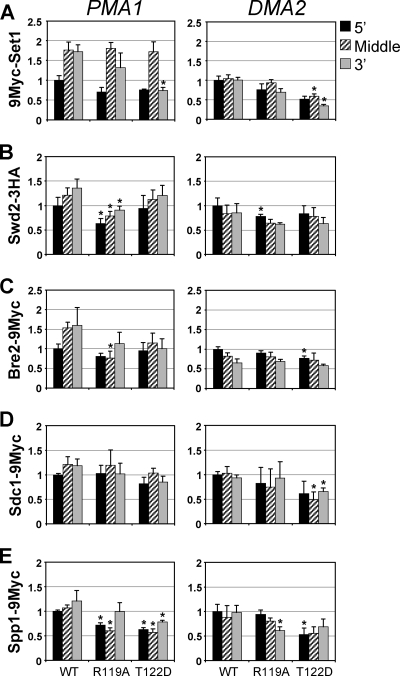
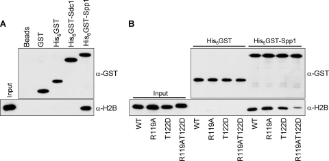
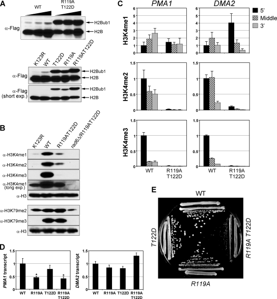
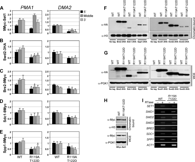
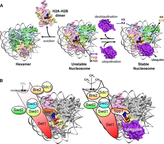
The trans-histone regulatory cross talk between H2BK123 ubiquitination (H2Bub1) and H3K4 and H3K79 methylation is not fully understood. In this study, we report that the residues arginine 119 and threonine 122 in the H2B C-terminal helix are important for transcription and cell growth and play a direct role in controlling H2Bub1 and H3K4 methylation. These residues modulate H2Bub1 levels by controlling the chromatin binding and activities of the deubiquitinases. Furthermore, we find an uncoupling of the H2Bub1-mediated coregulation of both H3K4 and -K79 methylation, as these H2B C-terminal helix residues are part of a distinct surface that affects only Set1-COMPASS (complex proteins associated with Set1)-mediated H3K4 methylation without affecting the functions of Dot1. Importantly, we also find that these residues interact with Spp1 and control the chromatin association, integrity, and overall stability of Set1-COMPASS independent of H2Bub1. Therefore, we have uncovered a novel role for the H2B C-terminal helix in the trans-histone cross talk as a binding surface for Set1-COMPASS. We provide further insight into the trans-histone cross talk and propose that H2Bub1 stabilizes the nucleosome by preventing H2A-H2B eviction and, thereby, retains the "docking site" for Set1-COMPASS on chromatin to maintain its stable chromatin association, complex stability, and processive methylation.


Figures

FIG. 1.
FIG. 1.
Presence of the residues following H2B residue K123 but not their primary sequence is needed for H2Bub1 and H3K4 methylation. (A) Amino acid sequence of the yeast histone H2B C-terminal region from valine (position 114) to alanine (position 130) is shown. Consensus sumoylation sites (in boldface) inserted to replace T122 and K123 in H2B to create H2B(2SU) and the two lysine-to-leucine mutations in H2B(2SU) (K1L and K2L) are shown. The sequence of mutant H2B with alanine substitutions or a truncation following K123 is also shown. Except for the control (H2B [no tag]), H2B and its derivatives contain a N-terminal Flag epitope. (B) Changes in sumoylation (su)/ubiquitination (ub1) and H3K4 methylation were assessed by Western blotting using antibodies that detect the Flag epitope (anti-Flag) or specific forms of H3K4 methylation (me1, monomethylation; me2, dimethylation; me3, trimethylation), respectively. (C and D) Western blots for H2Bub1 and H3K4 methylation, as described in panel B. A triangle represents a 2-fold serial dilution of the extract, a circle denotes a protein that cross-reacts with anti-Flag, and an asterisk denotes a cross-reacting antigen detected by some batches of anti-H3K4me1 (compare panels B and D). α, anti.
FIG. 2.
FIG. 2.
H2B C-terminal residues R119 and T122 control H2Bub1 levels, H3K4 methylation, and telomeric gene silencing. (A and D) Western blots for H2Bub1, as described in the legend to Fig. 1B. (B and E) Western blots for H3K4 and -K79 methylation. (C) Ribbon representations of the H2B C-terminal helix generated in the UCSF Chimera package using coordinates from the solved yeast nucleosome structure (Protein Data Bank [PDB] accession no. 1ID3) (54). Relevant amino acids are shown in ball and stick format. The diagonal white line demarcates the two phases of the helix. (F) Tenfold serial dilutions of yeast cultures were spotted on synthetic complete medium (SC) or medium-containing 5FOA and grown at 30°C for 2 days. WT, wild type.
FIG. 3.
FIG. 3.
H2B-R119A and H2B-T122D increase H2Bub1 levels by affecting deubiquitination. (A) Western blot for H2Bub1, as described in the legend to Fig. 1B. (B) Western blot analysis of Ubp8-3Flag, Rad6-9Myc, and Ubp10-3HA in the whole-cell extracts (WCE) and chromatin fractions detected using the indicated antibodies. The levels of Pgk1 and H3 serve as loading controls. (C) In vitro deubiquitination using whole-cell extracts obtained from the indicated ubp8Δ ubp10Δ strains mixed with either a mock extract or an extract from a wild-type strain containing both deubiquitinases and incubated for the indicated time. H2Bub1 levels were assessed as described for panel A.
FIG. 4.
FIG. 4.
H2B-R119A and H2B-T122D increase H2Bub1 levels and alter H3K4 methylation levels on transcribed genes. (A) Schematic diagrams of the gene organization for PMA1 and DMA2 are shown, drawn to scale. Solid black lines represent the 5′, middle (M), or 3′ ORF regions amplified by PCR. (B) Distribution and H2Bub1 occupancy were assessed by ChDIP assay using anti-Flag and an antibody that recognizes ubiquitinated proteins. Error bars show standard errors of the means obtained from two independent experiments. (C to E) Distribution and occupancy of H3K4 methylation was assessed by ChIP assays, and the occupancy for each modification was normalized to H3 occupancy. To show the change in distribution and overall occupancy of a histone modification across an ORF, the normalized occupancy obtained for all regions in the wild type and mutants are shown relative to the normalized occupancy obtained for the 5′ ORF region in the wild type (set to 1). Error bars show standard errors of the means obtained from three independent experiments.
FIG. 5.
FIG. 5.
H2B-R119A and H2B-T122D adversely affect the functions of normal and hyperactive Set1 in an H2Bub1-independent manner. (A to D) Western blots for H3K4 methylation in the indicated strains. Wild-type Set1 or its dominant, hyperactive form (Set1D-G990E) was expressed in the indicated strains containing wild type or mutant H2B (B), additionally lacking RAD6 (rad6Δ) (C), and strains lacking one of the Set1-COMPASS subunits SWD1, SDC1, or SPP1 (D). An asterisk denotes a cross-reacting antigen detected by some batches of anti-H3K4me1.
FIG. 6.
FIG. 6.
H2B-R119A and H2B-T122D reduce chromatin-bound levels of Sdc1 and Spp1. Whole-cell extracts (A to F) and the chromatin fraction (G to L) obtained from indicated strains were used for Western blotting to determine changes in the levels of the Set1-COMPASS subunits. The levels of Pgk1 and H3 serve as loading controls.
FIG. 7.
FIG. 7.
H2B-R119A and H2B-T122D affect the occupancy of Set1-COMPASS subunits on transcribed genes. (A to E) Distribution and occupancy of the indicated Set1-COMPASS subunits in 5′, middle, and 3′ ORF regions of PMA1 and DMA2 in the indicated strains determined by ChIP assay, as described in the legend to Fig. 4. Error bars show standard errors of the means obtained from at least three independent experiments. An asterisk denotes a statistically significant difference in factor occupancy at a given region between the mutant and the wild type, as determined by Student's t test (P < 0.05).
FIG. 8.
FIG. 8.
H2B C-terminal helix residues R119 and T122 are necessary for the interaction of Spp1 with H2B. (A) A bacterial lysate expressing H2B was incubated with purified GST, His6GST, His6GST-Spp1, or His6GST-Sdc1 before binding to glutathione-Sepharose beads. (B) Bacterial lysates expressing wild-type or mutant H2B were incubated with equal amounts of His6GST or His6GST-Spp1 (15 μM) prior to glutathione-Sepharose binding. Bead-bound H2B and GST-tagged proteins were detected using anti-H2B and anti-GST. A fraction of the bacterial lysate used in binding was probed with anti-H2B to evaluate the amount of soluble H2B (2%; input).
FIG. 9.
FIG. 9.
H2B-R119AT122D causes drastic changes in H2Bub1, H3K4 methylation, and gene expression levels and affects cell growth. (A to B) Western blots for H2Bub1, H3K4, and -K79 methylation in the indicated strains. Short exp., short exposure; long exp., long exposure. (C) ChIP assays for distribution and occupancy of H3K4 methylation were done as described in the legend to Fig. 4. (D) Transcript levels of PMA1 and DMA2 relative to the control ACT1 were measured by quantitative PCR using mRNA obtained from the indicated strains following reverse transcription. Changes in normalized transcript levels in the mutants are shown relative to that in the wild type, set to 1. Error bars show standard errors of the means obtained from three biological replicates. An asterisk indicates statistical significance (P < 0.05). (E) Cell growth of the indicated yeast strains was evaluated on rich medium at 30°C.
FIG. 10.
FIG. 10.
H2B-R119AT122D affects chromatin association and overall stability of Set1-COMPASS. (A to E) ChIP assay for the distribution and occupancy of Set1-COMPASS subunits in PMA1 and DMA2 in the indicated strains was done as described in the legends to Fig. 4 and 7. An asterisk shows statistical significance (P < 0.05). (F to H) Chromatin fractions or whole-cell extracts from indicated strains were analyzed to determine changes in the levels of Set1-COMPASS subunits. (I) Transcripts for Set1-COMPASS subunits were detected using reverse transcriptase PCR (RT-PCR) with the indicated strains. The levels of ACT1 serve as controls. RTase, reverse transcriptase; +, with reverse transcriptase in the RT-PCR; −, without reverse transcriptase in the RT-PCR. A representative of three independent experiments is shown.
FIG. 11.
FIG. 11.
Trans-histone regulation of H3K4 and -K79 methylation by H2Bub1. (A) Model for the dynamic changes in yeast nucleosome stability. Location of the residues involved in Set1-COMPASS docking (H2B residues R119 and T122), site of H2B ubiquitination (K123), and H3 residues modified by methylation (K4 and K79) are shown on the yeast nucleosome. C-terminal G76 of ubiquitin (PDB accession no. 1UBQ) is placed close to H2B residue K123 to simulate H2Bub1. (B) Speculative model depicting the role of H2Bub1 and the H2B C-terminal helix in modulating the chromatin association, integrity, and stability of Set1-COMPASS. Dotted lines denote weak intermolecular interactions, subunit dissociation, and unfavorable conformation. CH3 (gray), low levels of H3K4me1; CH3 (black), high levels of H3K4me1, K4me2, and K4me3. See the text for details.

Similar articles

Cited by

References

    1. Amati, B. B., and S. M. Gasser. 1988. Chromosomal ARS and CEN elements bind specifically to the yeast nuclear scaffold. Cell 54:967-978. - PubMed
    1. Berger, S. L. 2007. The complex language of chromatin regulation during transcription. Nature 447:407-412. - PubMed
    1. Briggs, S. D., T. Xiao, Z. W. Sun, J. A. Caldwell, J. Shabanowitz, D. F. Hunt, C. D. Allis, and B. D. Strahl. 2002. Gene silencing: trans-histone regulatory pathway in chromatin. Nature 418:498. - PubMed
    1. Chandrasekharan, M. B., F. Huang, and Z. W. Sun. 2009. Ubiquitination of histone H2B regulates chromatin dynamics by enhancing nucleosome stability. Proc. Natl. Acad. Sci. U. S. A. 106:16686-16691. - PMC - PubMed
    1. Dehé, P. M., M. Pamblanco, P. Luciano, R. Lebrun, D. Moinier, R. Sendra, A. Verreault, V. Tordera, and V. Géli. 2005. Histone H3 lysine 4 mono-methylation does not require ubiquitination of histone H2B. J. Mol. Biol. 353:477-484. - PubMed

Publication types

MeSH terms