. 2010 May 10;207(5):1081-93.
doi: 10.1084/jem.20092243. Epub 2010 May 3.

Inhibition of T cell response to native low-density lipoprotein reduces atherosclerosis

Affiliations

Inhibition of T cell response to native low-density lipoprotein reduces atherosclerosis

Andreas Hermansson et al. J Exp Med. .

Abstract

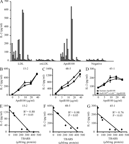
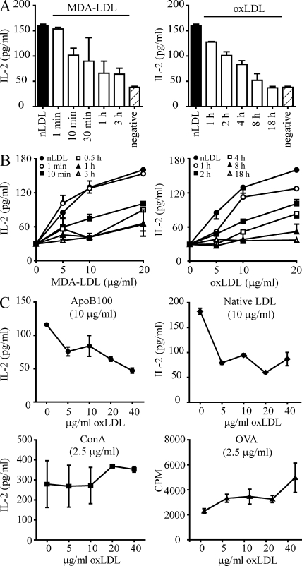
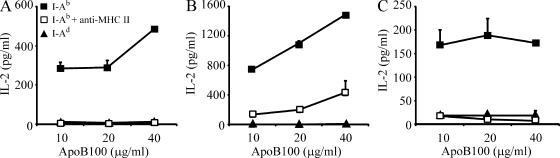
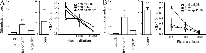
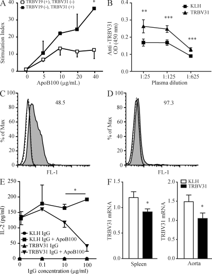
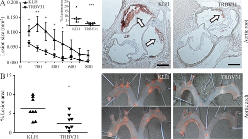
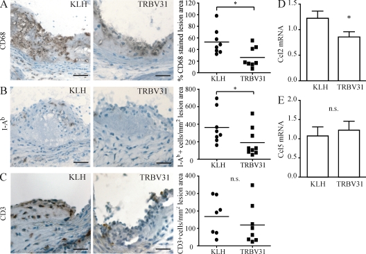
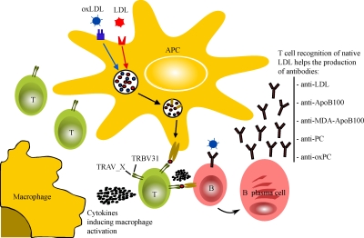
Immune responses to oxidized low-density lipoprotein (oxLDL) are proposed to be important in atherosclerosis. To identify the mechanisms of recognition that govern T cell responses to LDL particles, we generated T cell hybridomas from human ApoB100 transgenic (huB100(tg)) mice that were immunized with human oxLDL. Surprisingly, none of the hybridomas responded to oxidized LDL, only to native LDL and the purified LDL apolipoprotein ApoB100. However, sera from immunized mice contained IgG antibodies to oxLDL, suggesting that T cell responses to native ApoB100 help B cells making antibodies to oxLDL. ApoB100 responding CD4(+) T cell hybridomas were MHC class II-restricted and expressed a single T cell receptor (TCR) variable (V) beta chain, TRBV31, with different Valpha chains. Immunization of huB100(tg)xLdlr(-/-) mice with a TRBV31-derived peptide induced anti-TRBV31 antibodies that blocked T cell recognition of ApoB100. This treatment significantly reduced atherosclerosis by 65%, with a concomitant reduction of macrophage infiltration and MHC class II expression in lesions. In conclusion, CD4(+) T cells recognize epitopes on native ApoB100 protein, this response is associated with a limited set of clonotypic TCRs, and blocking TCR-dependent antigen recognition by these T cells protects against atherosclerosis.


Figures

Figure 1.
Figure 1.
T cell hybridomas recognize native LDL and its ApoB100 protein. (A) 105 hybridoma cells from each of 23 different clones were incubated with 4 × 10⁵ irradiated APCs with 20 µg /ml of LDL, oxLDL, or ApoB100. Each column represents one clone. Media without LDL were used as negative controls. Results are from one experiment. (B–D) 105 hybridoma cells of three different clones (B, hybridoma 15–2; C, 48–5; D, 45–1) were incubated with 4 × 10⁵ irradiated APCs and different concentrations of purified human ApoB100 from plasma LDL, or transgenic human ApoB100 from huB100tg x Ldlr−/− mice. Hybridomas 15–2, 48–5, and 45–1 (B–D) recognize purified and transgenic ApoB100 in a dose-dependent manner. In all experiments, IL-2 secretion was used as a measure of activation. Data show means ± SEM. Results are representative of three independent experiments. (E–G) 105 hybridoma cells were incubated with 4 × 10⁵ irradiated APCs with 20 µg/ml of LDL or LDL oxidized to different extents (copper oxidation [20 µM CuSO4] for varying lengths of time). After 24 h of incubation with the different preparations, IL-2 secretion was measured in the supernatant. X axis shows the mean of TBARS values and y axis shows the mean of IL-2 levels. All T cell hybridomas showed an inverse correlation between IL-2 levels and the degree of LDL oxidation. Results are representative of three independent experiments.
Figure 2.
Figure 2.
T cell recognition of LDL is decreased by oxidative modification. 105 hybridoma cells (48–5) were coincubated with 4 × 10⁵ irradiated APCs. (A) 20 µg/ml of LDL or LDL modified to different extents (malondialdehyde [MDA] or copper oxidation [20 µM CuSO4] for varying lengths of time) was added to the coincubation, and 24 h later IL-2 secretion was measured in the supernatant. Filled columns show response to native LDL and striped columns show response to medium control. (B) Dose–response curves for the coincubation with different concentrations of LDL-modified either with MDA or copper. (C) Co-incubation with fixed concentrations of ApoB100, native LDL, or concanavalin A together with increasing concentration of copper-oxidized LDL. For OVA, co-incubations were done with splenocytes from OT-II mice and activation was measured as CPM from [3H]thymidine incorporation, as described in the Materials and methods section. Data show means ± SEM. Results are representative of three independent experiments.
Figure 3.
Figure 3.
T cell responses to ApoB100 are I-Ab–restricted. 105 hybridoma cells were incubated with 4 × 10⁵ irradiated APCs from mice of either the I-Ab or I-Ad haplotype and different concentrations of human ApoB100. Hybridoma cells were also challenged with ApoB100 in the presence of a blocking antibody to MHC class II (I-Ab). After 24 h of incubation, IL-2 secretion was measured in the supernatant. A, 15–2 clone; B, 48–5 clone; C, 45–1 clone. Data show means ± SEM. Results are from one experiment.
Figure 4.
Figure 4.
Immunization with oxLDL or ApoB100 expands T cell populations that recognize native epitopes of LDL and induces antibodies to oxLDL, native LDL, and ApoB100. huB100tg x Ldlr−/− mice were immunized with oxLDL (A) or ApoB100 (B). 5 × 10⁵ splenocytes were challenged in vitro with 20 μg/mL of human oxLDL or native human ApoB100. Error bars represent the mean ± SEM of the stimulation index from [3H]thymidine incorporation, as described in the Materials and methods. Sera were assayed for antibodies to oxLDL, native LDL, and ApoB100. Values are expressed as mean ± SEM of the OD values for each dilution. Data are representative of two independent experiments (n = 4 mice per group). **, P < 0.01.
Figure 5.
Figure 5.
TRBV31+ T cells recognize ApoB100. (A) Depletion of TRBV31+ cells reduces T cell responses to ApoB100. HuB100tg x Ldlr−/− mice were immunized with ApoB100. Splenocytes, harvested after a booster injection, were depleted of TRBV31+ or TRBV19+ cells by fluorescence-activated cell sorting. 5 × 10⁵ TRBV31+/TRBV19 or TRBV19+/TRBV31 cells were challenged in vitro with different concentrations of human ApoB100. T cell activation was measured by [3H]thymidine incorporation and stimulation index, calculated as described in the Materials and methods section. Data show means ± SEM and are representative of two experiments. *, P < 0.05. (B) Induction of IgG antibodies to TRBV31 peptide after immunization with TRBV31. HuB100tg x Ldlr−/− mice were immunized and boosted with TRBV31 peptide conjugated to KLH, or with KLH alone as control. Serum IgG antibodies to TRBV31 peptide were measured by ELISA. Results are presented as mean ± SEM and representative of two independent experiments. **, P < 0.01; ***, P < 0.001. (C) Anti-TRBV31 IgG binds to LDL-reactive T cell hybridoma. Affinity-purified IgG antibodies from TRBV31-immunized mice were incubated with an LDL-reactive TRBV31+ hybridoma (48–5 clone), followed by incubation with FITC-labeled anti–mouse IgG and flow cytometric analysis. The graph shows representative histograms from 48–5 cells in the FITC channel (MFI, x axis) after labeling with IgG from mice immunized with the TRBV31-KLH conjugate (gray profile) or KLH alone (dashed line). Total IgG from untreated mice was used as negative control (black line). Results are representative of three independent experiments. (D) Anti-TRBV31 IgG does not bind to a non LDL-reactive T cell hybridoma. Affinity-purified IgG antibodies from TRBV31-immunized mice were incubated with a non–LDL-reactive hybridoma (97–3 clone), followed by incubation with FITC-labeled anti–mouse IgG and flow cytometric analysis. The graph shows representative histograms from 97–3 cells in the FITC channel (MFI, x axis) after labeling with IgG from mice immunized with the TRBV31-KLH (gray profile) conjugate (dashed line) or KLH alone. Total IgG from untreated mice was used as negative control (black line). Results are representative of three independent experiments. (E) Anti-TRBV31 IgG inhibits T cell recognition of ApoB100. 104 48–5 hybridoma cells were challenged in vitro with 20 µg /ml ApoB100 in the presence of 4 × 10⁴ irradiated APCs with IgG from mice immunized with TRBV31-KLH or KLH alone. After 24 h of incubation, IL-2 secretion was measured in the supernatant. Results show means ± SEM and are representative of three independent experiments. *, P < 0.05. (F) Immunization against TRBV31 reduces TRBV31 TCR mRNA in spleen and aorta. mRNA transcript levels were evaluated in aorta and spleen from huB100tg x Ldlrl−/− mice immunized with TRBV31-KLH conjugate or KLH alone. Bar graphs represent the mean ± SEM of TRBV31 mRNA relative to HPRT mRNA/CD3 mRNA relative to HPRT mRNA (n = 9 mice per group). *, P < 0.05. Results were pooled from two experiments.
Figure 6.
Figure 6.
Immunization against TRBV31 reduces atherosclerosis. HuB100tg x Ldlr−/− mice were immunized with the TRBV31 peptide-KLH conjugate or with KLH alone. The mice were sacrificed after 10 wk on a Western diet. (A) Atherosclerotic lesion size in the proximal aorta. Lesions were analyzed by microscopic morphometry of serial sections at 100–800 µm from the aortic valves. Data show cross-sectional lesion size (mm2). Inset shows means of the eight cross-sectional lesion sizes for each animal, and the line indicates mean value per group of mice. n = 9 mice per group. Pooled data are presented from two independent experiments. *, P < 0.05; **, P < 0.01; ***, P < 0.001. Representative micrographs from immunized mice. Hematoxylin-Oil red O staining, original magnification 20×. White arrows indicate lesion areas. Bars, 250 µm. (B) Atherosclerotic lesion size in the aortic arch. Dissected arches were stained with Sudan IV en face and the percentage lesion area of total vessel area was calculated using ImageJ image analysis software. The additive area of all the plaques in a given aortic arch was calculated as a percentage of the total surface area of the arch. n = 8 mice per group. Two representative stained samples from each group are shown. *, P < 0.05.
Figure 7.
Figure 7.
Immunization against TRBV31 reduces inflammation in atherosclerotic lesions. (A) Reduced accumulation of macrophages in lesions of TRBV31-immunized mice. Images show CD68 immunostaining of representative aortic root sections from mice immunized with KLH alone (left) or the TRBV31-KLH conjugate (right). The dot plot shows proportion of total lesion area stained by CD68 antibody in the two groups. Data show values for individual mice and mean value for each group (line; n = 8 mice per group). (B) Reduced MHC class II (I-Ab) expression in lesions of TRBV31-immunized mice. Images show I-Ab immunostaining of representative sections from the aortic root. The dot plot shows the number of I-Ab+ cells per mm2 lesion area in the two groups (n = 8 mice for KLH and 9 mice for TRBV31). (C) No change in CD3 expression in lesions of TRBV31-immunized mice. Images show CD3 immunostaining of representative sections from the aortic root. The dot plot shows the number of CD3+ cells per square millimeter of lesion area in the two groups (n = 7 mice for KLH and 8 mice for TRBV31). n.s., not significant. Immunoperoxidase labeling, original magnification 100×. Bars: (A–C) 100 µm. (D–E) Immunization with TRBV31 reduces CCL2 mRNA. mRNA levels of CCL2 and CCL5 were measured by real-time RT-PCR in aorta from huB100tg x Ldlr−/− mice immunized with TRBV31-KLH conjugate or KLH alone. Error bars represent the mean ± SEM for mRNA under study/mRNA for the housekeeping gene, HPRT (n = 9 mice per group). *, P < 0.05 in A–D. Results in A–E show pooled data from two experiments.
Figure 8.
Figure 8.
Proposed hypothesis for CD4+ T cell recognition of LDL. LDL particles are taken up by LDL receptors and, if modified, by scavenger receptors on the APC. Uptake could also be mediated by Fc receptors ligating opsonized LDL particles. After endosomal degradation, peptides derived from ApoB100 are transferred into the antigen-presenting pathway, where they bind to nascent MHC class II molecules. The peptide–MHC complexes are transported to the cell surface and recognized by CD4+ T cells equipped with TRBV31+ TCR. The ensuing T cell activation leads to cytokine secretion that can promote macrophage activation and inflammation. Once T cells are activated toward native ApoB100 epitopes, they may provide help for B cells recognizing native LDL, ApoB100, or lipids such as phosphocholine (PC), but also oxidatively modified epitopes such as MDA-ApoB100 or oxPC. T, T cells; B, B cells; blue symbol, Scavenger receptors or Fc receptors; red symbol, LDL receptors.

References

    1. Ait-Oufella H., Salomon B.L., Potteaux S., Robertson A.K., Gourdy P., Zoll J., Merval R., Esposito B., Cohen J.L., Fisson S., et al. 2006. Natural regulatory T cells control the development of atherosclerosis in mice. Nat. Med. 12:178–180 10.1038/nm1343 - DOI - PubMed
    1. Andersson J., Libby P., Hansson G.K. 2010. Adaptive immunity and atherosclerosis. Clin. Immunol. 134:33–46 10.1016/j.clim.2009.07.002 - DOI - PubMed
    1. Berliner J.A., Heinecke J.W. 1996. The role of oxidized lipoproteins in atherogenesis. Free Radic. Biol. Med. 20:707–727 10.1016/0891-5849(95)02173-6 - DOI - PubMed
    1. Berliner J.A., Heinecke J.W. 1996. The role of oxidized lipoproteins in atherogenesis. Free Radic. Biol. Med. 20:707–727 10.1016/0891-5849(95)02173-6 - DOI - PubMed
    1. Binder C.J., Hartvigsen K., Chang M.K., Miller M., Broide D., Palinski W., Curtiss L.K., Corr M., Witztum J.L. 2004. IL-5 links adaptive and natural immunity specific for epitopes of oxidized LDL and protects from atherosclerosis. J. Clin. Invest. 114:427–437 - PMC - PubMed

Publication types

MeSH terms