. 2010 May;92(5):1088-96.
doi: 10.2106/JBJS.I.00686.

Location and initiation of degenerative rotator cuff tears: an analysis of three hundred and sixty shoulders

Affiliations

Location and initiation of degenerative rotator cuff tears: an analysis of three hundred and sixty shoulders

H Mike Kim et al. J Bone Joint Surg Am. 2010 May.

Abstract

Background: It has been theorized that degenerative rotator cuff tears most commonly involve the supraspinatus tendon, initiating at the anterior portion of the supraspinatus insertion and propagating posteriorly. The purposes of this study were to determine the most common location of degenerative rotator cuff tears and to examine tear location patterns associated with various tear sizes.

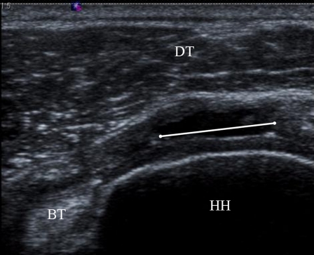
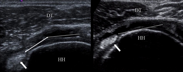
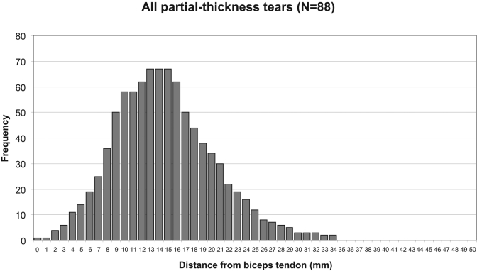
Methods: Ultrasonograms of 360 shoulders with either a full-thickness rotator cuff tear (272) or a partial-thickness rotator cuff tear (eighty-eight) were obtained to measure the width and length of the tear and the distance from the biceps tendon to the anterior margin of the tear. Tears were grouped on the basis of their size (anteroposterior width) and extent (partial or full-thickness). Each tear was represented numerically as a column of consecutive numbers representing the tear width and distance posterior to the biceps tendon. All tears were pooled to graphically represent the width and location of the tears within groups. Frequency histograms of the pooled data were generated, and the mode was determined for each histogram representing various tear groups.

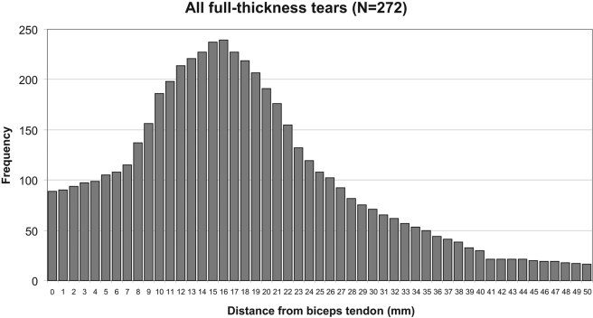
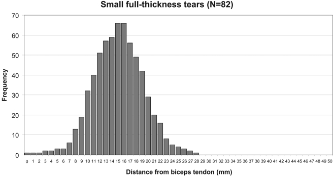
Results: The mean age (and standard deviation) of the 233 subjects (360 shoulders) was 64.7 +/- 10.2 years. The mean width and length of the tears were 16.3 +/- 12.1 mm and 17.0 +/- 13.0 mm, respectively. The mean distance from the biceps tendon to the anterior tear margin was 7.8 +/- 5.7 mm (range, 0 to 26 mm). Histograms of the various tear groups invariably showed the location of 15 to 16 mm posterior to the biceps tendon to be the most commonly torn location within the posterior cuff tendons. The histograms of small tears (a width of <10 mm) and partial-thickness tears showed similar distributions of tear locations, indicating that the region approximately 15 mm posterior to the biceps tendon may be where rotator cuff tears most commonly initiate.

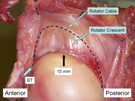
Conclusions: Degenerative rotator cuff tears most commonly involve a posterior location, near the junction of the supraspinatus and infraspinatus. The patterns of tear location across multiple tear sizes suggest that degenerative cuff tears may initiate in a region 13 to 17 mm posterior to the biceps tendon.


Figures

Fig. 1
Fig. 1
Measurement of the width of a fluid-filled full-thickness tear in the transverse plane. The width (white line) was defined as the distance between the anterior and posterior torn tendon ends. BT = biceps tendon, HH = humeral head, DT = deltoid muscle.
Fig. 2
Fig. 2
Measurement of the distance from the biceps tendon to the anterior margin of a tear in the transverse plane. A tear (dotted line) with an anterior margin that has a measurable distance (solid line) from the biceps (arrow) is shown on the left. The distance from the biceps to a fluid-filled full-thickness tear (dotted line) that extends up to the biceps tendon (arrow) is not measurable, as the rotator interval is disrupted (shown on the right). HH = humeral head, DT = deltoid muscle.
Fig. 3-A Fig. 3-B
Fig. 3-A Fig. 3-B
Fig. 3-A A histogram of all full-thickness tears shows a unimodal distribution. The most common location (i.e., the mode) is 16 mm posterior to the biceps, and the second most common location is 15 mm posterior to the biceps. The frequency represents the number of tears that involve a given location within the rotator cuff tendon. The location is shown as the distance in millimeters from the intra-articular portion of the biceps tendon. Fig. 3-B A histogram of the small full-thickness tears shows a unimodal distribution. The most common locations are 15 and 16 mm posterior to the biceps. The frequency represents the number of tears that involve a given location within the rotator cuff tendon. The location is shown as the distance in millimeters from the intra-articular portion of the biceps tendon.
Fig. 3-A Fig. 3-B
Fig. 3-A Fig. 3-B
Fig. 3-A A histogram of all full-thickness tears shows a unimodal distribution. The most common location (i.e., the mode) is 16 mm posterior to the biceps, and the second most common location is 15 mm posterior to the biceps. The frequency represents the number of tears that involve a given location within the rotator cuff tendon. The location is shown as the distance in millimeters from the intra-articular portion of the biceps tendon. Fig. 3-B A histogram of the small full-thickness tears shows a unimodal distribution. The most common locations are 15 and 16 mm posterior to the biceps. The frequency represents the number of tears that involve a given location within the rotator cuff tendon. The location is shown as the distance in millimeters from the intra-articular portion of the biceps tendon.
Fig. 4
Fig. 4
A histogram of all partial-thickness tears shows a unimodal distribution. The most common locations are 13, 14, and 15 mm posterior to the biceps. The frequency represents the number of tears that involve a given location within the rotator cuff tendon. The location is shown as the distance in millimeters from the intra-articular portion of the biceps tendon.
Fig. 5
Fig. 5
A cadaver shoulder specimen showing the articular surface of the rotator cuff and the humeral head. The rotator cable (curved dashed line) spans from the biceps tendon (BT) to the inferior border of the infraspinatus. Distal to the rotator cable is the rotator crescent. In this specimen, a mild degenerative change (a small defect in the synovium, black arrow) is seen on the articular surface of the rotator cuff at a location approximately 15 mm posterior to the biceps tendon.

References

    1. Matsen FA, III, Arntz CT, Lippitt SB. Rotator cuff. : The shoulder. Rockwood CA, Matsen FA, III, Philadelphia: W.B. Saunders; 1998
    1. Codman EA. The shoulder. Boston: Norman Publishing; 1934
    1. Codman EA, Akerson IB. The pathology associated with rupture of the supraspinatus tendon. Ann Surg. 1931;93:348-59 - PMC - PubMed
    1. Keyes EL. Observations on rupture of the supraspinatus tendon: based upon a study of seventy-three cadavers. Ann Surg. 1933;97:849-56 - PMC - PubMed
    1. Hijioka A, Suzuki K, Nakamura T, Hojo T. Degenerative change and rotator cuff tears. An anatomical study in 160 shoulders of 80 cadavers. Arch Orthop Trauma Surg. 1993;112:61-4 - PubMed

Publication types