. 2010 May 18;107(20):9299-304.
doi: 10.1073/pnas.1003909107. Epub 2010 May 3.

Pregnancy induces a fetal antigen-specific maternal T regulatory cell response that contributes to tolerance

Affiliations

Pregnancy induces a fetal antigen-specific maternal T regulatory cell response that contributes to tolerance

Daniel A Kahn et al. Proc Natl Acad Sci U S A. .

Abstract

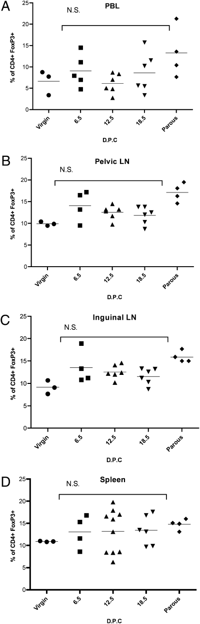
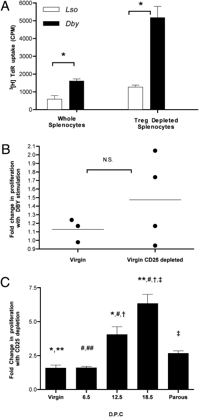
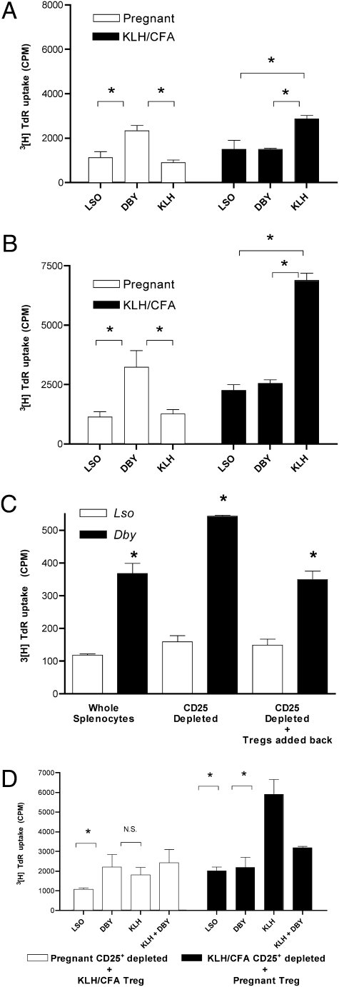
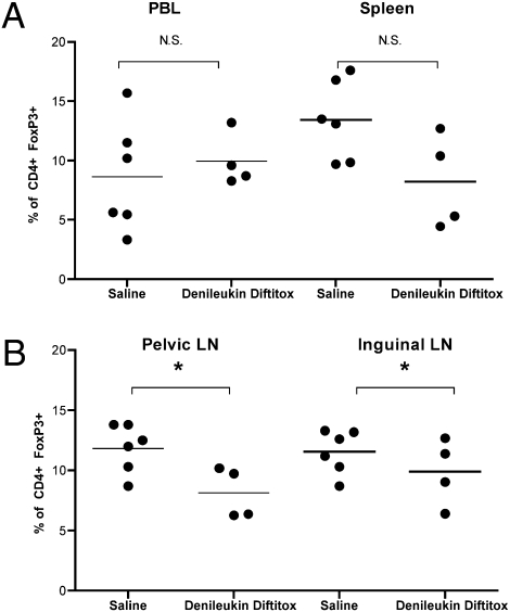
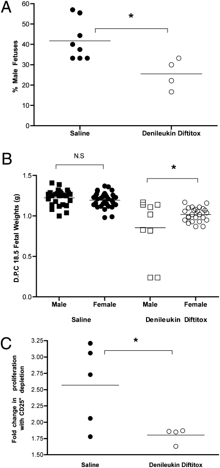
A fetus is inherently antigenic to its mother and yet is not rejected. The T regulatory (Treg) subset of CD4(+) T cells can limit immune responses and has been implicated in maternal tolerance of the fetus. Using virgin inbred mice undergoing a first syngenic pregnancy, in which only the male fetuses are antigenic, we demonstrate a maternal splenocyte proliferative response to the CD4(+) T cell restricted epitope of the male antigen (H-Y) in proportion to the fetal antigen load. A portion of the maternal immune response to fetal antigens is Treg in nature. The bystander suppressive function of pregnancy-generated Tregs requires the presence of the fetal antigen, demonstrating their inherent antigen specificity. In vivo targeting of diphtheria toxin to kill Tregs leads to a lower fraction of live male offspring and a selective reduction in mass of the surviving males. Thus, Tregs generated in the context of pregnancy function in an antigen-specific manner to limit the maternal immune response to the fetus in a successful pregnancy.


Conflict of interest statement

The authors declare no conflict of interest.

Figures

Fig. 1.
Fig. 1.
Pregnancy imparts CD4+ T cell awareness. (A) Splenocytes harvested from virgin (8 weeks old, n = 4) and previously bred (14 weeks old, n = 4) C57BL/6 mice from the same colony: Cellular proliferation assay performed using 1 × 10⁶ cells per mL to the CD4+ T cell peptide epitope of the H-Y antigen presented by I-Ab (10 μM Dby), the I-Ab presented lysteriolysin peptide epitope (10 μM Lso), or no peptide stimulation. Cultures were pulsed with 1 μCi per well 3[H]TdR for the final 18 h of a 72-h culture (cpm ± SD). Statistical difference determined by one-way ANOVA with Bonferroni–Dunn posttest (N.S., nonsignificant; *, P < 0.05). Pregnancy primes maternal CD4+ T cell awareness in proportion to fetal antigenic mass. (B) Splenocytes harvested from virgin 8-week-old C57BL/6 mice (8 weeks, n = 4) and timed first pregnancy of 8-week-old female C57BL/6 × C57BL/6 males (n = 4 each time point) at days post coitus (dpc 6.5, 12.5, 18.5): Cellular proliferation assay performed using 1 × 10⁶ cells per mL to the CD4+ T cell peptide epitope of the H-Y antigen presented by I-Ab (10 μM Dby) or the I-Ab presented lysteriolysin peptide epitope (10 μM Lso). Cultures were pulsed with 1 μCi per well 3[H]TdR for final 18 h of a 72-h culture. Composite fold increase in proliferation with Dby/Lso ± SEM. Statistical difference determined by one-way ANOVA with Bonferroni–Dunn posttest (*, **, #, P < 0.05). (C) Days postcoitus 6.5. Splenocyte proliferative response determined for individual mice ± SD along with fetal gender determination. (D) Days postcoitus 12.5. (E) Days postcoitus 18.5. Statistical difference determined by two-tailed, unpaired Student's t test (N.S., nonsignificant; *, P < 0.05).
Fig. 2.
Fig. 2.
Regional distribution of T regulatory cells in relation to first syngenic mating. Single-cell suspensions from indicated lymphoid organs prepared from virgin (n = 3) or timed first pregnancies from C57BL/6 syngenic mating (dpc 6.5, n = 4; dpc 12.5, n = 6; dpc 18.5, n = 6), or retired from syngenic breeding (n = 4). FACS performed with gating on CD4+ cells to determine the fraction of FoxP3+ cells. Statistical differences determined by one-way ANOVA with Bonferroni–Dunn posttest (N.S., nonsignificant).
Fig. 3.
Fig. 3.
Pregnancy results in a functional T regulatory response in proportion to the fetal mass. (A) Splenocytes harvested from a timed first pregnancy of a 6-week-old C57BL/6 × C57BL/6 male at dpc = 18.5 (six male fetuses). Splenocyte proliferative response determined for individual mice ± SD. (B) Splenocytes from virgin 6-week-old C57BL/6 (no prior plugs). Treg depletion by magnetic bead separation of CD25+ cells. Cellular proliferation assay was performed using 1 × 10⁶ cells per mL to the CD4+ T cell peptide epitope of the H-Y antigen presented by I-Ab (10 μM Dby) or the I-Ab presented lysteriolysin peptide epitope (10 μM Lso). Cultures were pulsed with 1μCi per well 3[H]TdR for the final 18 h of a 72-h culture (cpm ± SD). Statistical difference determined by two-tailed unpaired Student's t test (N.S., nonsignificant; *, P < 0.05). (C) Splenocytes from virgin, timed first pregnancy of C57BL/6 × C57BL/6 (8 weeks old, n = 4 each time point) dpc 6.5, 12.5, and 18.5, or 14-week-old retired breeder (parous). Depletion of CD25+ cells (Treg) by magnetic bead. Composite fold increase in proliferation with Dby (CD25+ depleted)/Dby (whole splenocytes) ± SEM. Statistical difference determined by one-way ANOVA with Bonferroni–Dunn posttest (*, **, #, †, ‡, P < 0.05).
Fig. 4.
Fig. 4.
Pregnancy-induced T regulatory cells suppressive function is antigen-specific. (A) Splenocytes harvested from a timed first pregnancy of 6-week-old C57BL/6 × C57BL/6 male at dpc 18.5 (four male fetuses) as well as virgin 6-week-old C57BL/6 female immunized with KLH/CFA 7 days prior. Cellular proliferation assay was performed using 1 × 10⁶ cells per mL to the CD4+ T cell peptide epitope of the H-Y antigen presented by I-Ab (10 μM Dby), the I-Ab presented lysteriolysin peptide epitope (10 μM Lso) or KLH (1 μg/mL). Cultures were pulsed with 1 μCi per well 3[H]TdR for the final 18 h of a 72-h culture. (B) CD25+ reduced splenocytes. (C) Splenocytes harvested from a timed first pregnancy of 6 weeks – old C57BL/6 x C57BL/6 male at dpc 15.5 (three male fetuses). CD25+ cell reduced. CD25+ cells (Treg) added back to CD25+-depleted splenocytes in a ratio of 4:1. (D) CD25+-reduced splenocytes from either the dpc 18.5 mouse (□) or the KLH/CFA-immunized mouse (■) above cocultured with the CD25+ (Treg enriched population) isolated by magnetic bead positive selection from the KLH/CFA-immunized mouse (□) or the pregnant mouse (■) at a ratio of 4:1. Stimuli as indicated (Lso 10 μM, Dby 10 μM, KLH 1 μg/mL). Statistical difference determined by one-way ANOVA with Bonferroni–Dunn posttest (N.S., nonsignificant; *, P < 0.05).
Fig. 5.
Fig. 5.
In vivo effect of denileukin diftitox on Treg populations in regional lymphoid organs. Shown is FACS analysis of indicated single-cell suspensions immediately ex vivo stained for CD4 and FoxP3 from timed first pregnancy of C57BL/6 × C57BL/6 at dpc 18.5. Saline, n = 6; denileukin diftitox, n = 4. Statistical difference was determined by two-tailed, unpaired Student's t test (N.S., nonsignificant; *, P < 0.05).
Fig. 6.
Fig. 6.
In vivo reduction in Tregs results in poor pregnancy outcome specific to fetal antigens. (A) Percentage male fetuses determined for each litter of C57BL/6 × C57BL/6 by PCR from Saline-treated (●) (n = 8) or denileukin diftitox–treated (○) mice (n = 4). (B) On dpc 18.5, fetal weights determined for fetuses from the above mating. (C) Splenocytes harvested from the saline-treated (●) (n = 5) or denileukin diftitox–treated (○) (n = 4) mice, and cellular proliferation assay performed using 1 × 10⁶ cells per mL to the CD4+ T cell peptide epitope of the H-Y antigen presented by I-Ab (10 μM Dby) or the I-Ab presented lysteriolysin peptide epitope (10 μM Lso). Cultures were pulsed with 1 μCi per well 3[H]TdR for the final 18 h of a 72-h culture. Depletion of CD25+ cells (Treg) by magnetic bead. Composite fold increase in proliferation with Dby (CD25+ depleted)/Dby (whole splenocytes). Statistical difference was determined by two-tailed, unpaired Student's t test (N.S., nonsignificant; *, P < 0.05).

References

    1. Billingham RE, Brent L, Medawar PB. Actively acquired tolerance of foreign cells. Nature. 1953;172:603–606. - PubMed
    1. Trowsdale J, Betz AG. Mother's little helpers: Mechanisms of maternal-fetal tolerance. Nat Immunol. 2006;7:241–246. - PubMed
    1. Mold JE, et al. Maternal alloantigens promote the development of tolerogenic fetal regulatory T cells in utero. Science. 2008;322:1562–1565. - PMC - PubMed
    1. Barinaga M. Cells exchanged during pregnancy live on. Science. 2002;296:2169–2172. - PubMed
    1. Niederkorn JY. See no evil, hear no evil, do no evil: The lessons of immune privilege. Nat Immunol. 2006;7:354–359. - PubMed

Publication types