. 2010 May 18;107(20):9452-7.
doi: 10.1073/pnas.1000675107. Epub 2010 May 3.

A domain swap approach reveals a role of the plant wall-associated kinase 1 (WAK1) as a receptor of oligogalacturonides

Affiliations

A domain swap approach reveals a role of the plant wall-associated kinase 1 (WAK1) as a receptor of oligogalacturonides

Alexandre Brutus et al. Proc Natl Acad Sci U S A. .

Abstract

Oligogalacturonides (OGs) released from the plant cell wall are active both as damage-associated molecular patterns (DAMPs) for the activation of the plant immune response and regulators of plant growth and development. Members of the Wall-Associated Kinase (WAK) family are candidate receptors of OGs, due to their ability to bind in vitro these oligosaccharides. Because lethality and redundancy have hampered the study of WAKs by reverse genetics, we have adopted a chimeric receptor approach to elucidate the role of Arabidopsis WAK1. In a test-of-concept study, we first defined the appropriate chimera design and demonstrated that the Arabidopsis pattern recognition receptor (PRR) EFR is amenable to the construction of functional and resistance-conferring chimeric receptors carrying the ectodomain of another Arabidopsis PRR, FLS2. After, we analyzed chimeras derived from EFR and WAK1. Our results show that, upon stimulation with OGs, the WAK1 ectodomain is capable of activating the EFR kinase domain. On the other hand, upon stimulation with the cognate ligand elf18, the EFR ectodomain activates the WAK1 kinase, triggering defense responses that mirror those normally activated by OGs and are effective against fungal and bacterial pathogens. Finally, we show that transgenic plants overexpressing WAK1 are more resistant to Botrytis cinerea.


Conflict of interest statement

The authors declare no conflict of interest.

Figures
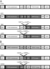
Fig. 1.
Fig. 1.
Constructs for the expression of the chimeric receptors. (A) The coding regions of EFR and FLS2 are labeled in white and gray, respectively, with the region corresponding to the signal peptide for translocation into the ER indicated in black. eJM and iJM, external and internal juxtamembrane region, respectively; TM, transmembrane region. (B) The coding regions of EFR and WAK1 are labeled in dark and light gray, respectively, with the region corresponding to the signal peptides indicated in black. All genes were fused to the GFP-encoding sequence and placed under the control of the CaMV 35S promoter. NOS: nos terminator. Annotated amino acids indicate the junction points in the two chimeric receptors.
Fig. 2.
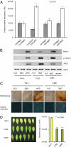
Fig. 2.
Functional characterization of EFR/FLS2 chimeric receptors. (A) Induction of ethylene biosynthesis in agroinfiltrated tobacco leaves expressing the indicated fluorescent receptors. Excised infiltrated leaf sectors were stimulated for 2 h with the elicitor (10 μM elf18 or 1 μM flg22) as indicated in the figure. Agroinfiltrated leaf tissues expressing fluorescent EFR or FLS2 and elicited with flg22 or elf18, respectively, represented our negative control. Values are means ± SEM (n = 3). (B) Elicitor-induced accumulation of Ret-Ox, CYP81F2, and PAD3 transcripts analyzed by semiquantitative PCR, using the UBQ5 gene for normalization, in transgenic Arabidopsis ecotype Ws-0 plants stably expressing eJMC. Leaves were incubated with H2O, flg22 (1 μM), elf18 (10 μM), or a generic peptide (10 μM) for 30 min. The experiment was repeated three times with similar results. The same experiment was performed in a second independent transgenic line expressing eJMC with similar results. (C) Histochemical analysis of defense responses induced upon 1 h of treatment with H2O, flg22 (1 μM), or elf18 (10 μM) in transgenic Arabidopsis ecotype Ws-0 plants stably expressing eJMC. (Upper) H2O2 accumulation revealed by 3,3’-diaminobenzidine tetrahydrochloride staining. (Lower) Callose deposition revealed by aniline blue staining. (D) Infections by spray-inoculation with Pseudomonas syringae pv. tomato (Pst) DC3000 of transgenic Arabidopsis ecotype Ws-0 plants stably expressing eJMC. (Left) Disease symptoms of wild-type and transformed plants at 4 days postinfection (dpi). (Right) Growth of Pst at 3 dpi. Higher resistance of eJMC plants, compared to Ws-0 untransformed plants, was found in three independent experiments. No difference was observed when infections were performed by syringe-infiltration.
Fig. 3.
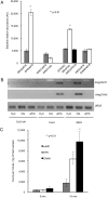
Fig. 3.
Induction of defense responses by WAK1/EFR chimeric receptors. (A) Induction of ethylene biosynthesis in agroinfiltrated tobacco explants expressing EFR, WAK1, WEG, and EWAK. Explants were stimulated for 2 h with elf18 (10 μM), OGs (100 μg/mL), or short and biologically inactive OGs (OG3-6). Agroinfiltrated leaf tissues expressing EFR or WEG and elicited with OGs or elf18 respectively, represented our negative control. Values are means ± SEM (n = 3). (B) Elicitor-induced gene expression in untransformed Col-0 and Col-0 efr plants and in transgenic Arabidopsis Col-0 efr plants stably expressing WEG. Accumulation of At3g22270 and At4g37640 transcripts was analyzed by semiquantitative PCR, using the UBQ5 gene for normalization. Adult leaves were syringe-infiltrated with water, OGs (25 μg/mL), or elf18 (10 μM) for 3 h. The experiment was repeated three times with similar results. The same experiment was performed in a second independent transgenic line expressing WEG with similar results. (C) Oxidative burst in agroinfiltrated tobacco leaves expressing EWAK. Leaves expressing eJMC and EFR were used as a negative and a positive control, respectively. The burst was measured by photon counting using leaf slices incubated in a solution containing 10 μM elf18, luminol, and peroxidase. The experiment was repeated at least six times with two independent replicates.
Fig. 4.
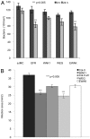
Fig. 4.
Response to bacterial and fungal pathogens of plants expressing the chimeric receptors. (A) Growth of A. tumefaciens in Nicotiana tabacum tissues transiently expressing receptor proteins. Tobacco leaves were infiltrated with Agrobacterium carrying the indicated receptor constructs and the number of Agrobacterium cfu in the tissues was analyzed immediately after infiltration (black bars) and after 24 h (gray bars). EFR and eJMC were used as positive and negative controls, respectively. For each receptor, three samples were analyzed in five independent experiments. Asterisks indicate statistically significant differences against control (Col-0 for WAK1 plants and Col-0 efr for WEG and EWAK plants). (B) Lesion development in Arabidopsis wild-type (Col-0) and mutant Col-0 efr plants and in transgenic plants expressing WAK1, WEG, and EWAK plants inoculated with B. cinerea at 48 h postinoculation. Values are means ± SEM of at least 16 lesions. The same experiment was performed in a second independent transgenic line expressing WAK1 with similar results. Asterisks indicate statistically significant differences against control (Col-0 for WAK1 plants and Col-0 efr for WEG and EWAK plants). No symptoms were observed in plants inoculated with Potato Dextrose Broth (PDB) (mock-inoculation).

References

    1. Jiang D, Liang J, Noble PW. Hyaluronan in tissue injury and repair. Annu Rev Cell Dev Biol. 2007;23:435–461. - PubMed
    1. Puré E, Assoian RK. Rheostatic signaling by CD44 and hyaluronan. Cell Signal. 2009;21:651–655. - PMC - PubMed
    1. Scheibner KA, et al. Hyaluronan fragments act as an endogenous danger signal by engaging TLR2. J Immunol. 2006;177:1272–1281. - PubMed
    1. Willats WG, McCartney L, Mackie W, Knox JP. Pectin: Cell biology and prospects for functional analysis. Plant Mol Biol. 2001;47:9–27. - PubMed
    1. Ridley BL, O'Neill MA, Mohnen D. Pectins: Structure, biosynthesis, and oligogalacturonide-related signaling. Phytochemistry. 2001;57:929–967. - PubMed

Publication types

MeSH terms