. 2010 May 25;107(21):9590-5.
doi: 10.1073/pnas.1004104107. Epub 2010 May 3.

Electrophilic tuning of the chemoprotective natural product sulforaphane

Affiliations

Electrophilic tuning of the chemoprotective natural product sulforaphane

Young-Hoon Ahn et al. Proc Natl Acad Sci U S A. .

Abstract

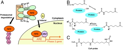
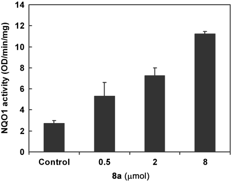
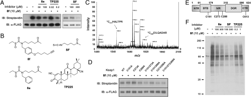
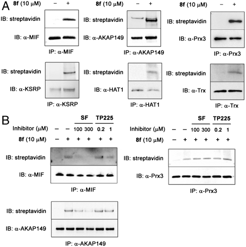
Sulforaphane [1-isothiocyanato-4-(methylsulfinyl)butane], a naturally occurring isothiocyanate derived from cruciferous vegetables, is a highly potent inducer of phase 2 cytoprotective enzymes and can protect against electrophiles including carcinogens, oxidative stress, and inflammation. The mechanism of action of sulforaphane is believed to involve modifications of critical cysteine residues of Keap1, which lead to stabilization of Nrf2 to activate the antioxidant response element of phase 2 enzymes. However, the dithiocarbamate functional group formed by a reversible reaction between isothiocyanate of sulforaphane and sulfhydryl nucleophiles of Keap1 is kinetically labile, and such modification in intact cells has not yet been demonstrated. Here we designed sulforaphane analogs with replacement of the reactive isothiocyanate by the more gentle electrophilic sulfoxythiocarbamate group that also selectively targets cysteine residues in proteins but forms stable thiocarbamate adducts. Twenty-four sulfoxythiocarbamate analogs were synthesized that retain the structural features important for high potency in sulforaphane analogs: the sulfoxide or keto group and its appropriate distance to electrophilic functional group. Evaluation in various cell lines including hepatoma cells, retinal pigment epithelial cells, and keratinocytes as well as in mouse skin shows that these analogs maintain high potency and efficacy for phase 2 enzyme induction as well as the inhibitory effect on lipopolysaccharide-induced nitric oxide formation like sulforaphane. We further show in living cells that a sulfoxythiocarbamate analog can label Keap1 on several key cysteine residues as well as other cellular proteins offering new insights into the mechanism of chemoprotection.


Conflict of interest statement

The authors declare no conflict of interest.

Figures

Fig. 1.
Fig. 1.
Proposed chemoprotective mechanism of sulforaphane for phase 2 gene activation involves covalent Cys modification of Keap1. (A) Mechanism of phase 2 enzyme induction. In the basal state, the transcription factor Nrf2 is efficiently ubiquitylated and targeted for proteasomal degradation by forming a complex with Keap1 and Cul3 E3 ligase. Inducers such as sulforaphane modify cysteines of Keap1, which leads to stabilization and translocation of Nrf2 to the nucleus, and its binding to ARE and stimulation of phase 2 gene transcription. (B) Reactions of sulforaphane and sulfoxythiocarbamate analog with a thiol. The isothiocyanate group of sulforaphane reacts with sulfhydryl groups on protein to form dithiocarbamate adduct that appear to be kinetically labile (Top), whereas sulfoxythiocarbamate analog forms a relatively stable thiocarbamate derivative (Bottom). (C) Structure of sulfoxythiocarbamate CoA probe developed previously (24).
Fig. 2.
Fig. 2.
Structures of sulfoxythiocarbamate sulforaphane analogs. Two classes of analogs, one with sulfoxide group (4a–4r) and another with ketone-group (8a–8f), were generated with alkyl or aryl modifications on R1 and R2 positions.
Fig. 3.
Fig. 3.
Evaluation of sulfoxythiocarbamate analog for NQO1 enzyme induction on mouse skin. The back of each SKH-1 hairless mouse (n = 3) was topically treated with three concentrations of 8a (indicated amounts in 40 μL of 80% aq. acetone) and solvent only over ca. 2.5 cm2 area for three doses at 24-h intervals. Mice were euthanized 24 h after the last dose, and dorsal skin was harvested. NQO1 specific activity was measured in supernatant fractions of homogenates of skin sections treated with 8a or solvent (control). Means ± SD are shown.
Fig. 4.
Fig. 4.
Sulfoxythiocarbamate analog conjugates on Keap1 cysteines as well as potential target proteins in cells. (A) Labeling FLAG-Keap1 in cells. HEK293 cells transiently expressing FLAG-tagged Keap1 were treated with 8f for 0.5 h, or pretreated with 8a, sulforaphane, or TP225 for 0.5 h before incubation of 8f for 0.5 h. FLAG-Keap1 was immunoprecipitated from cell lysates with anti-FLAG antibody, subjected to reaction with biotin azide and eluted with FLAG peptide. Samples were immunoblotted with streptavidin (Top) and anti-FLAG antibody (Bottom). (B) Structure of compounds, 8f, 8a, sulforaphane, and TP225. (C) MALDI-MS spectrum of FLAG-Keap1 tryptic digests labeled by 8f. Eluted FLAG-Keap1 was digested by trypsin, followed by incubation with avidin-coated beads. Biotin-conjugated peptides were eluted with aq. acetonitrile and analyzed by MALDI mass spectrometry, which showed two peaks corresponding to modified Cys 273 and Cys 288 peptides. (D) Labeling FLAG-Keap1 cysteine to alanine mutants. Cells transiently expressing FLAG-tagged WT or mutant Keap1 were treated with 8f for 0.5 h. FLAG-Keap1 proteins, eluted after immunoprecipitation with anti-FLAG antibody and conjugation with biotin azide, were immunoblotted with streptavidin (Top) and anti-FLAG antibody (Bottom). (E) Schematic structure of Keap1 with five domains that include NTR, BTB, IVR, DGR, and CTR. Cys 288 and Cys 613 modified peptides were found by LTQ mass analysis. (F) Labeling potential target proteins in cells. After incubation of compounds as described in A, cell lysates were subjected to click reaction with biotin azide, and analyzed by SDS-PAGE and immunoblotting with streptavidin HRP.
Fig. 5.
Fig. 5.
Validation of selected target proteins labeled by sulfoxythiocarbamate analog 8f in cells. (A) Target proteins labeled by 8f. HEK293 cells were treated with 8f or vehicle for 2.5 h. (B) Competition by sulforaphane or TP225. HEK293 cells were pretreated with sulforaphane, TP225, or vehicle for 0.5 h before incubation with 8f for 0.5 h. Each protein was immunoprecipitated from cell lysates, subjected to click reaction with biotin azide on beads. Eluted samples were immunoblotted with streptavidin as well as protein specific antibodies.

References

    1. Talalay P, Fahey JW. Phytochemicals from cruciferous plants protect against cancer by modulating carcinogen metabolism. J Nutr. 2001;131:3027S–3033S. - PubMed
    1. Surh YJ. Cancer chemoprevention with dietary phytochemicals. Nat Rev Cancer. 2003;3:768–780. - PubMed
    1. Zhang Y, Talalay P, Cho CG, Posner GH. A major inducer of anticarcinogenic protective enzymes from broccoli–isolation and elucidation of structure. Proc Natl Acad Sci USA. 1992;89:2399–2403. - PMC - PubMed
    1. Juge N, Mithen RF, Traka M. Molecular basis for chemoprevention by sulforaphane: A comprehensive review. Cell Mol Life Sci. 2007;64:1105–1127. - PMC - PubMed
    1. Zhang Y, et al. Anticarcinogenic activities of sulforaphane and structurally related synthetic norbornyl isothiocyanates. Proc Natl Acad Sci USA. 1994;91:3147–3150. - PMC - PubMed

Publication types

MeSH terms

LinkOut - more resources