. 2010 May 18;107(20):9176-81.
doi: 10.1073/pnas.0912295107. Epub 2010 May 3.

Structure of the EF-hand domain of polycystin-2 suggests a mechanism for Ca2+-dependent regulation of polycystin-2 channel activity

Affiliations

Structure of the EF-hand domain of polycystin-2 suggests a mechanism for Ca2+-dependent regulation of polycystin-2 channel activity

Edward T Petri et al. Proc Natl Acad Sci U S A. .

Abstract

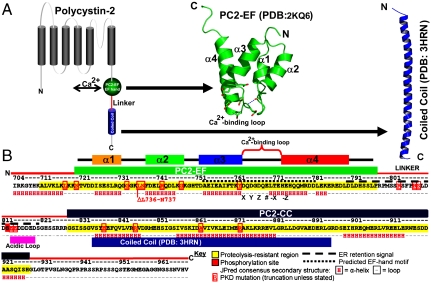
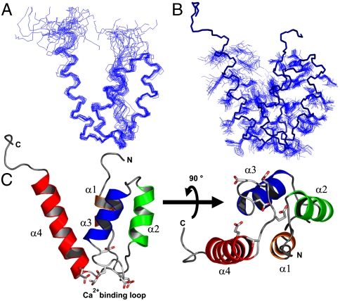
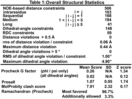
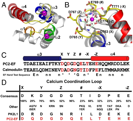
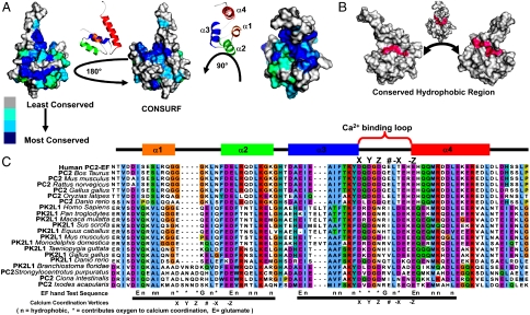
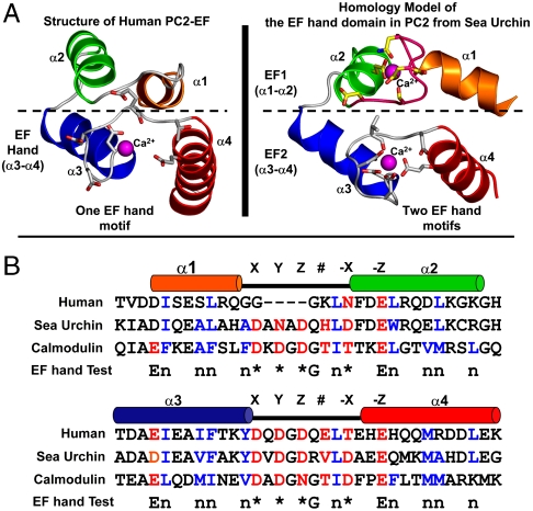
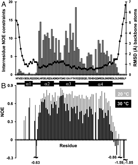
The C-terminal cytoplasmic tail of polycystin-2 (PC2/TRPP2), a Ca(2+)-permeable channel, is frequently mutated or truncated in autosomal dominant polycystic kidney disease. We have previously shown that this tail consists of three functional regions: an EF-hand domain (PC2-EF, 720-797), a flexible linker (798-827), and an oligomeric coiled coil domain (828-895). We found that PC2-EF binds Ca(2+) at a single site and undergoes Ca(2+)-dependent conformational changes, suggesting it is an essential element of Ca(2+)-sensitive regulation of PC2 activity. Here we describe the NMR structure and dynamics of Ca(2+)-bound PC2-EF. Human PC2-EF contains a divergent non-Ca(2+)-binding helix-loop-helix (HLH) motif packed against a canonical Ca(2+)-binding EF-hand motif. This HLH motif may have evolved from a canonical EF-hand found in invertebrate PC2 homologs. Temperature-dependent steady-state NOE experiments and NMR R(1) and R(2) relaxation rates correlate with increased molecular motion in the EF-hand, possibly due to exchange between apo and Ca(2+)-bound states, consistent with a role for PC2-EF as a Ca(2+)-sensitive regulator. Structure-based sequence conservation analysis reveals a conserved hydrophobic surface in the same region, which may mediate Ca(2+)-dependent protein interactions. We propose that Ca(2+)-sensing by PC2-EF is responsible for the cooperative nature of PC2 channel activation and inhibition. Based on our results, we present a mechanism of regulation of the Ca(2+) dependence of PC2 channel activity by PC2-EF.


Conflict of interest statement

The authors declare no conflict of interest.

Figures

Fig. 1.
Fig. 1.
Molecular architecture of Polycystin-2. (A) PC2 contains three functional regions in its C-terminal tail: PC2-EF (green sphere and NMR structure) which undergoes Ca2+-dependent conformational changes, a linker (red), and oligomeric coiled coil (blue cylinder and cartoon crystal structure). (B) Sequence of the PC2 C-terminal tail: PC2-EF (green), linker (red), and coiled coil (PC2-CC, blue). Helices α1–4 in PC2-EF are orange, green, blue, and red bars. Residues in the coiled coil X-ray structure are shown as a blue bar (8). Ca2+-binding residues are labeled X, Y, Z, #, -X, and -Z. ADPKD mutations (http://pkdb.mayo.edu) are highlighted (red boxes).
Fig. 2.
Fig. 2.
Structure of Ca2+-bound PC2-EF. (A) Backbone ensemble structure of PC2-EF. The top 20 conformers were aligned (backbone rmsd 0.6 Å over I725-D790) (blue ribbons). (B) Backbone atoms for the top model (heavy blue ribbon) and side chain atoms for the top 20 conformers (blue lines) C) Overall structure of PC2-EF. Helices α1–4 (orange, green, blue, and red), and Ca2+-coordinating residues [Corey-Pauling-Koltun (CPK) sticks] are shown.
Table 1
Table 1
Z scores are with respect to a standard set of ∼250 X-ray structures (17).
Fig. 3.
Fig. 3.
Comparison of PC2-EF with CaM. (A) Structural alignment of PC2-EF (cartoon, helices α1–4 colored orange, green, blue, and red, and loops colored yellow) with Ca2+-bound CaM (PDB ID: 1CLL, white cartoon, magenta Ca2+ spheres). (B) The PC2-EF Ca2+-binding loop aligned with CaM. CaM Ca2+-coordinating residues are labeled (X, Y, Z, #, -X, -Z, white CPK sticks, magenta Ca2+ sphere). Ca2+-coordination bonds are shown as dashes. The residue # coordinates Ca2+ with a backbone carbonyl, and -X coordinates Ca2+ through an ordered water. Analogous X, Y, Z, #, -X, -Z residues in PC2-EF are shown as yellow CPK sticks. (C) Sequence alignment of the EF-hand from PC2-EF and CaM (PC2-EF helices α3 and α4, blue and red). X, Y, Z, #, -X, -Z residues are red and labeled. (D) Consensus sequence in EF-hand Ca2+-binding loops (adapted from ref. 19) at X, Y, Z, #, -X, -Z positions. Sequence similarity (%) and common residue substitutions are shown. The Ca2+-binding loop sequence of PC2-EF (red) and polycystin-L (PK2L1, black) is shown.
Fig. 4.
Fig. 4.
Sequence conservation and conserved surface analysis of PC2-EF. (A) PC2-EF surface conservation (blue = most conserved) from CONSURF. PC2-EF contains a conserved surface in a V-shaped cleft opposite the Ca2+-binding site formed by α1, α3, and α4. (B) Hydrophobic saddle in PC2-EF wrapping the conserved surface. (C) CLUSTALW alignment of PC2-EF with EF-hands from the C-terminal tails of PC2 and polycystin-L from vertebrates and invertebrates (colored by CLUSTALX conservation). A test sequence corresponding to consensus EF-hands is shown.
Fig. 5.
Fig. 5.
Comparison of human PC2-EF with a homology model of sea urchin PC2-EF. (A) Homology model of sea urchin PC2-EF (Right) constructed using Ca2+-bound CaM (PDB ID: 1CLL) as a template in Swiss-Modeller (http://swissmodel.expasy.org/) compared with human PC2-EF (Left). Helices corresponding to human PC2-EF α1, α2, α3, and α4 are colored orange, green, blue, and red. Four residues deleted in the α1–α2 loop of human PC2-EF are colored pink in the sea urchin PC2-EF model. Ca2+ ions shown (magenta spheres) are from Ca2+-bound CaM. (B) Sequence alignment of human and sea urchin PC2-EF with CaM. Helices corresponding to human PC2-EF helices α1 through α4 are colored orange, green, blue, and red. Ca2+-coordinating residues in CaM are labeled X, Y, Z, #, -X, -Z, and colored red. Hydrophobic residues conserved in canonical EF-hands are colored blue. A test sequence corresponding to a consensus EF-hand (18) is shown.
Fig. 6.
Fig. 6.
Interresidue NOE restraints and backbone rmsds compared to steady-state NOE values. (A) Number of interresidue NOE constraints (gray bar chart, left axis) per residue overlaid with backbone rmsd per residue calculated as deviation from a mean structure of the top 20 conformers (right axis, scatter plot, black dots). (B) Steady-state NOE analysis at 20 °C (gray bars) and 30 °C (black bars) per residue.

Similar articles

Cited by

References

    1. Wu G, et al. Somatic inactivation of Pkd2 results in polycystic kidney disease. Cell. 1998;93(2):177–188. - PubMed
    1. Torres VE, Harris PC. Autosomal dominant polycystic kidney disease: The last 3 years. Kidney Int. 2009;76(2):149–168. - PMC - PubMed
    1. Nauli SM, et al. Polycystins 1 and 2 mediate mechanosensation in the primary cilium of kidney cells. Nat Genet. 2003;33(2):129–137. - PubMed
    1. Koulen P, et al. Polycystin-2 is an intracellular calcium release channel. Nat Cell Biol. 2002;4(3):191–197. - PubMed
    1. Qian F, et al. PKD1 interacts with PKD2 through a probable coiled-coil domain. Nat Genet. 1997;16(2):179–183. - PubMed

Publication types

MeSH terms