. 2010 May 18;107(20):9099-104.
doi: 10.1073/pnas.1004220107. Epub 2010 May 3.

Direct role for the RNA polymerase domain of T7 primase in primer delivery

Affiliations

Direct role for the RNA polymerase domain of T7 primase in primer delivery

Bin Zhu et al. Proc Natl Acad Sci U S A. .

Abstract

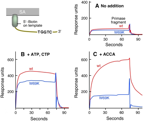
Gene 4 protein (gp4) encoded by bacteriophage T7 contains a C-terminal helicase and an N-terminal primase domain. After synthesis of tetraribonucleotides, gp4 must transfer them to the polymerase for use as primers to initiate DNA synthesis. In vivo gp4 exists in two molecular weight forms, a 56-kDa form and the full-length 63-kDa form. The 56-kDa gp4 lacks the N-terminal Cys(4) zinc-binding motif important in the recognition of primase sites in DNA. The 56-kDa gp4 is defective in primer synthesis but delivers a wider range of primers to initiate DNA synthesis compared to the 63-kDa gp4. Suppressors exist that enable the 56-kDa gp4 to support the growth of T7 phage lacking gene 4 (T7Delta4). We have identified 56-kDa DNA primases defective in primer delivery by screening for their ability to support growth of T7Delta4 phage in the presence of this suppressor. Trp69 is critical for primer delivery. Replacement of Trp69 with lysine in either the 56- or 63-kDa gp4 results in defective primer delivery with other functions unaffected. DNA primase harboring lysine at position 69 fails to stabilize the primer on DNA. Thus, a primase subdomain not directly involved in primer synthesis is involved in primer delivery. The stabilization of the primer by DNA primase is necessary for DNA polymerase to initiate synthesis.


Conflict of interest statement

The authors declare no conflict of interest.

Figures

Fig. 1.
Fig. 1.
Gene 4 proteins of bacteriophage T7. (A) Structure of T7 DNA primase [PDB ID code 1NUI, (6)]. The primase of T7 gp4 contains three major parts: the ZBD (magenta), the TOPRIM subdomain (blue), and the N-terminal subdomain of RPD (yellow). The latter two are collectively designated the RPD. Metal atoms are presented as silver dots, and tryptophan 69 is highlighted in red. (B) Schematic representation of the 56- and 63 kDa gp4. The primase is located in the N-terminal half of gp4, and the helicase is in the C-terminal half.
Fig. 2.
Fig. 2.
Delivery of oligonucleotides by gp4 to DNA polymerase during coordinated DNA synthesis. Coordinated DNA synthesis was carried out as described previously (22, 23). The reaction mixture contained 40 mM Tris-HCl (pH 7.5), 10 mM MgCl2, 10 mM DTT, 100 mg/mL bovine serum albumin, 50 mM potassium glutamate, 600 μM each dATP, dCTP, dGTP, and dTTP, 100 nM minicircle, 80 nM T7 DNA polymerase, 60 nM gp4 (63-kDa gp4 or 56-kDa gp4 as indicated) monomer (10 nM gp4 hexamer), and 4 μM gp2.5. Incorporation of [3H] dGMP into the leading strand (open symbols) or [3H] dCMP into the lagging strand (solid symbols) was measured and plotted against time. (A) Coordinated DNA synthesis in the presence of 300 μM each ATP and CTP. (B) 150 μM oligoribonucleotide r(ACCA) replaced ATP and CTP in the reaction in A. (C) 150 μM oligoribonucleotide r(GGGACCA) replaced ATP and CTP in the reaction in A. (D) Coordinated DNA synthesis supported by T7 63-kDa or 56-kDa gene 4 protein in the presence of various primers. The rates of leading- and lagging-strand synthesis and the ratio of lagging-strand synthesis to leading-strand synthesis are listed. The oligonucleotides and their complementarity to the lagging strand template (some of them to the primase recognition sequence 5′-TGGTC-3′) are presented. Final concentration of all the performed primers is 150 μM and E. coli total tRNA is 6 μM in the assay. Data were obtained from duplicate experiments.
Fig. 3.
Fig. 3.
Effect of alteration of tryptophan 69 on functions of 63-kDa gp4. (A) DNA-dependent dTTP hydrolysis activity of 63-kDa gp4. The indicated amounts of wild-type gp4 and gp4 with amino acid substitutions for Trp69 were incubated with 0.25 mM [α-32P] dTTP (0.1 μCi) in the presence of 8 nM M13 ssDNA at 37 °C for 20 min. Reaction products were separated by TLC, and dTTP hydrolysis was determined by measuring the amount of dTDP present in the reaction. (B) Template-directed primer synthesis of 63-kDa gp4. Reaction mixtures containing 10 μM template DNA, 5′-d(GGGTCA10), 0.1 mM each of ATP and [α-32P] CTP (0.1 μCi), and various amounts of the indicated gp4 were incubated at 37 °C for 30 min, and the reaction products were analyzed on denaturing polyacrylamide gels. The amount of CMP incorporated into tetraribonucleotide r(ACCC) was measured. (C and D) RNA-primed DNA synthesis in the presence of NTPs (C) or preformed primer r(ACCA) (D). Reaction mixtures containing 10 nM M13 ssDNA, 0.1 mM all four NTPs or 25 μM r(ACCA) as indicated, 0.3 mM all four dNTPs, 0.1 μCi of [α-32P] dGTP, 100 nM T7 DNA polymerase (a 1∶1 complex of T7 gene 5 protein and E. coli thioredoxin), and various amounts of the indicated gp4 were incubated at 37 °C for 10 min. Amounts of dGMP incorporated into DNA were measured and plotted against the concentration of gp4 used. Data were obtained from triplicate experiments.
Fig. 4.
Fig. 4.
Substitution of lysine for tryptophan 69 of gp4. (A) DNA unwinding activity was determined by measuring the amount of radiolabeled ssDNA displaced from a minireplication fork (500 fmol) by the indicated gp4. (B) Template-directed oligoribonucleotide synthesis catalyzed by 63-kDa gp4. Reaction mixtures containing 20 μM 15mer DNA 5′-d(GGGTCA10) or 10 nM M13 ssDNA, 0.1 mM each of [α-32P] CTP (0.1 μCi) and ATP (replaced with 0.1 mM each of four NTPs plus 0.5 mM dTTP when M13 ssDNA was used as template), and 22, 67, or 200 nM gp4 were incubated at 37 °C for 30 min, and the reaction products were analyzed on denaturing polyacrylamide gels. (C) The amount of CMP incorporated into tetraribonucleotide as shown in B with M13 ssDNA template was measured. (D) Coordinated DNA synthesis supported by 63-kDa gp4-W69K or 56-kDa gp4-W69K. Reactions were carried out as described for Fig. 3. The reaction mixture contained 40 mM Tris-HCl (pH 7.5), 10 mM MgCl2, 10 mM DTT, 100 mg/mL bovine serum albumin, 50 mM potassium glutamate, 600 μM each dATP, dCTP, dGTP, and dTTP, 100 nM minicircle, 80 nM T7 DNA polymerase, 60 nM gp4 monomer (10 nM gp4 hexamer), 4 μM gp2.5, and 300 μM each ATP and CTP for 63-kDa gp4-W69K or 150 μM r(ACCA) for 56-kDa gp4-W69K. Incorporation of [3H] dGMP into the leading strand (▪ for 63-kDa gp4-W69K and □ for 56-kDa gp4-W69K) or [3H] dCMP into the lagging strand (• for 63-kDa gp4-W69K and ◯ for 56-kDa gp4-W69K) was measured and plotted against time. Data were obtained from at least duplicate experiments.
Fig. 5.
Fig. 5.
Sequence-specific DNA binding of T7 primase. A biotinylated 25-mer ssDNA containing a primase recognition site, 5′-d(TGGTC) (1000 RU) was immobilized on the surface of a Biacore streptavidin sensor chip. T7 primase (10 μM) was flowed over the chip together with the indicated components in a buffer containing 40 mM Tris-HCl, pH 7.5, 50 mM potassium glutamate, 10 mM MgCl2, and 1 mM DTT, and binding signals were detected. Concentrations of ATP/CTP and r(ACCA) were 50 μM. Representative data from multiple experiments are presented.
Fig. 6.
Fig. 6.
Model for primer delivery by T7 primase. (A) Structure of T7 primase [PDB ID code 1NUI, (6)]. The ZBD is colored in red, the TOPRIM subdomain in blue, the N-terminal subdomain of RPD in yellow. Metal atoms are presented as silver dots. (B) Model of the predelivery complex formed by the 63-kDa gp4 (helicase not shown), DNA template (purple), and RNA primer. The 3′ end of the annealed oligonucleotide is stabilized by Trp69 in the N-terminal subdomain of the RPD while the other end of the primer/template is sandwiched by the ZBD and the TOPRIM subdomain. The 3′ end of the primer (5′ end of template) is ready to be extruded through the N-terminal subdomain of the RPD. (C) Model of the predelivery complex formed by the 56-kDa gp4 (helicase not shown), DNA template (purple), and RNA primer. The overall arrangement is similar as that for 63-kDa gp4. However, in the absence of the ZBD, longer oligonucleotides with noncomplementary 5′ termini can be accommodated. The 3′ end of the primer must match the template sequence and is stabilized by Trp69 as for the 63-kDa gp4. (D) Structure of the RPD domain of E. coli primase [PDB ID codes 1DD9 and 1DDE, (30)]. The TOPRIM subdomain is colored in blue and the N-terminal subdomain of RPD in yellow. (E) Schematic presentation of the complex (15) of E. coli primase with a short ssDNA from which the predelivery complex of T7 primase was derived. In both primases the tryptophan residues discussed in the text are highlighted.

Similar articles

Cited by

References

    1. Frick DN, Richardson CC. DNA primases. Annu Rev Biochem. 2001;70:39–80. - PubMed
    1. Ahnert P, Picha KM, Patel SS. A ring-opening mechanism for DNA binding in the central channel of the T7 helicase-primase protein. EMBO J. 2000;19:3418–3427. - PMC - PubMed
    1. Hamdan SM, Richardson CC. Motors, switches, and contacts in the replisome. Annu Rev Biochem. 2009;78:205–243. - PubMed
    1. Hamdan SM, Loparo JJ, Takahashi M, Richardson CC, van Oijen AM. Dynamics of DNA replication loops reveal temporal control of lagging-strand synthesis. Nature. 2009;457:336–339. - PMC - PubMed
    1. Lee JB, et al. DNA primase acts as a molecular brake in DNA replication. Nature. 2006;439:621–624. - PubMed

Publication types

MeSH terms

LinkOut - more resources