. 2010 May 18;107(20):9111-6.
doi: 10.1073/pnas.0914300107. Epub 2010 May 3.

Cotranslational structure acquisition of nascent polypeptides monitored by NMR spectroscopy

Affiliations

Cotranslational structure acquisition of nascent polypeptides monitored by NMR spectroscopy

Cédric Eichmann et al. Proc Natl Acad Sci U S A. .

Abstract

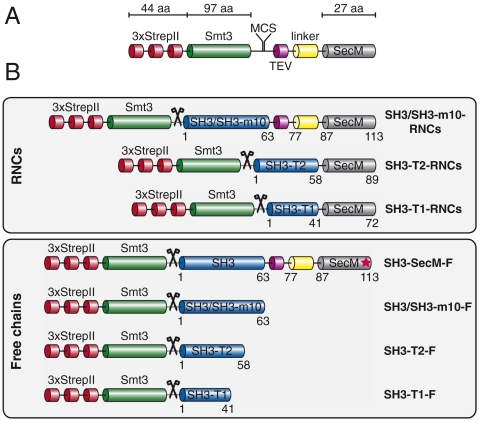
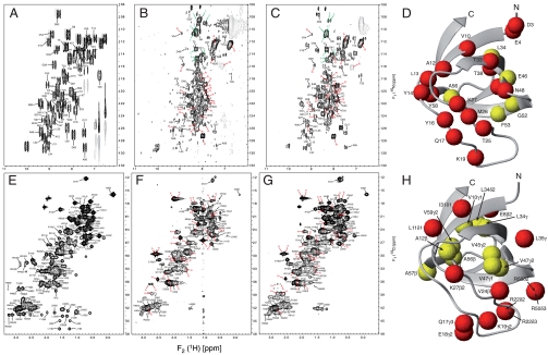
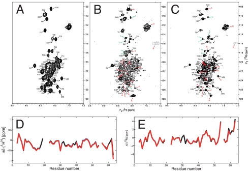
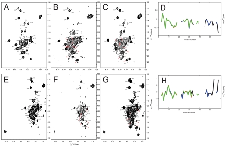
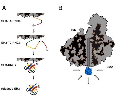
The folding of proteins in living cells may start during their synthesis when the polypeptides emerge gradually at the ribosomal exit tunnel. However, our current understanding of cotranslational folding processes at the atomic level is limited. We employed NMR spectroscopy to monitor the conformation of the SH3 domain from alpha-spectrin at sequential stages of elongation via in vivo ribosome-arrested (15)N,(13)C-labeled nascent polypeptides. These nascent chains exposed either the entire SH3 domain or C-terminally truncated segments thereof, thus providing snapshots of the translation process. We show that nascent SH3 polypeptides remain unstructured during elongation but fold into a compact, native-like beta-sheet assembly when the entire sequence information is available. Moreover, the ribosome neither imposes major conformational constraints nor significantly interacts with exposed unfolded nascent SH3 domain moieties. Our data provide evidence for a domainwise folding of the SH3 domain on ribosomes without significant population of folding intermediates. The domain follows a thermodynamically favorable pathway in which sequential folding units are stabilized, thus avoiding kinetic traps during the process of cotranslational folding.


Conflict of interest statement

The authors declare no conflict of interest.

Figures

Fig. 1.
Fig. 1.
Schematic representation of arrested RNCs and other constructs used for NMR analysis. (A) The plasmid for production of RNCs consists of an N-terminal triple StrepII tag (Red) followed by a Smt3 domain (Green), a multiple cloning site (MCS), a recognition sequence for the TEV protease (Magenta), a flexible linker (Yellow), and the SecM stalling peptide (Dark Gray). (B) Arrested RNCs and free polypeptides investigated by NMR. All constructs were expressed with an Ulp1-cleavable N-terminal triple StrepII-Smt3 tag, which had been removed in the final constructs subjected to NMR measurements (indicated by scissors representing the cleavage point). The numbers indicate the amino acid positions from the N terminus after Ulp1 treatment. A mutation in the SecM sequence (R163A; numbering according to the native SecM sequence) leads to loss of translational arrest and release of the polypeptide chain from the ribosome (Red Asterisk).
Fig. 2.
Fig. 2.
SH3 folds on ribosomes. [15N,1H]-CRINEPT-HMQC spectra of (A) SH3-F, (B) SH3-RNCs, (C) SH3 cleaved from the ribosome. [13C,1H]-HMQC spectra of (E) SH3-F, (F) SH3-RNCs, (G) SH3 cleaved from the ribosome. The peaks are labeled with the single amino acid code for SH3 in black, a red R for background signals emanating presumably from the ribosome, and a green L for residues assigned to the linker region added to the construct. (D and H) The observed NMR probes (Spheres) are spread over the entire SH3 domain. 15N-1H-moieties are shown in D and 13C-1H-moieties are shown in H. Red spheres indicate those cross peaks that exhibited line broadening due to nonspecific, transient interactions between the SH3 domain and the ribosome. Yellow spheres indicate a lack of line broadening of the cross peaks of interest.
Fig. 3.
Fig. 3.
Nascent SH3-m10 has a random coil-like conformation. [15N,1H]-CRINEPT-HMQC spectra of (A) SH3-m10-F, (B) SH3-m10-RNCs, and (C) SH3-m10 cleaved from the ribosome. Peak labels are as in Fig. 2. (D) 1H and (E) 15N chemical shift deviations from the corresponding random coil chemical shifts of SH3-m10-RNCs (Blue), SH3-m10 cleaved from the ribosome (Red) and SH3-m10-F free in solution (Black) are plotted versus the amino acid sequence.
Fig. 4.
Fig. 4.
Nascent SH3-T1 and SH3-T2 are unfolded. [15N,1H]-CRINEPT-HMQC spectra of (A) SH3-T1-F, (B) SH3-T1-RNCs, and (C) SH3-T1-F mixed with ribosomes. [15N,1H]-CRINEPT-HMQC spectra of (E) SH3-T2-F, (F) SH3-T2-RNCs, and (G) SH3-T2-F mixed with ribosomes. Peak labels are as in Fig. 2. (D) 1H and (H) 15N chemical shift deviations from the corresponding random coil chemical shifts of SH3-T1-RNCs (Red), SH3-T1-F free in solution (Green), SH3-T2-RNCs (Blue) and SH3-T2-F (Black) are shown versus the amino acid sequence.
Fig. 5.
Fig. 5.
Model of the domainwise cotranslational folding of nascent SH3. (A) Schematic model: The SH3 domain remains unfolded until the entire domain is synthesized and exposed outside of the ribosomal tunnel. (B) A scale model of cotranslationally folded native SH3 domain (Blue) tethered to the large ribosomal subunit (Gray, cut in half to visualize the tunnel interior) demonstrating the shielding capacity of the ribosome. The arrows indicate the hemisphere of the nascent polypeptide that would be accessible to interactions with cytosolic factors.

Similar articles

Cited by

References

    1. Wegrzyn RD, Deuerling E. Molecular guardians for newborn proteins: ribosome-associated chaperones and their role in protein folding. Cell Mol Life Sci. 2005;62:2727–2738. - PMC - PubMed
    1. Ban N, et al. The complete atomic structure of the large ribosomal subunit at 2.4 Å resolution. Science. 2000;289:905–920. - PubMed
    1. Yusupov MM, et al. Crystal structure of the ribosome at 5.5 Å resolution. Science. 2001;292:883–896. - PubMed
    1. Ramakrishnan V. Ribosome structure and the mechanism of translation. Cell. 2002;108:557–572. - PubMed
    1. Seidelt B, et al. Structural insight into nascent polypeptide chain-mediated translational stalling. Science. 2009;326:1412–1415. - PMC - PubMed

Publication types