. 2010 Jul;185(3):1111-28.
doi: 10.1534/genetics.110.117531. Epub 2010 May 3.

Functional genomics analysis of the Saccharomyces cerevisiae iron responsive transcription factor Aft1 reveals iron-independent functions

Affiliations

Functional genomics analysis of the Saccharomyces cerevisiae iron responsive transcription factor Aft1 reveals iron-independent functions

Sharon Berthelet et al. Genetics. 2010 Jul.

Abstract

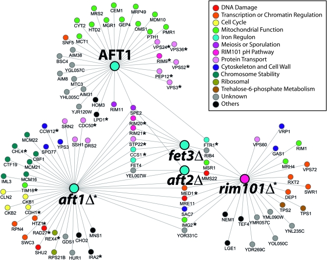
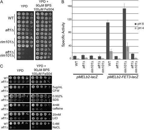
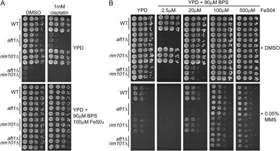
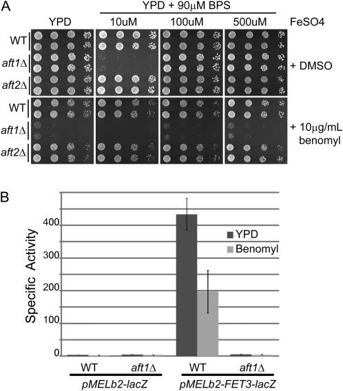
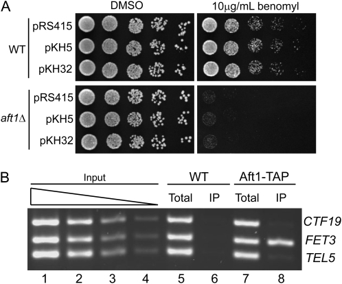
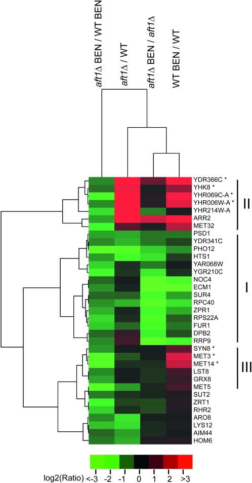
The Saccharomyces cerevisiae transcription factor Aft1 is activated in iron-deficient cells to induce the expression of iron regulon genes, which coordinate the increase of iron uptake and remodel cellular metabolism to survive low-iron conditions. In addition, Aft1 has been implicated in numerous cellular processes including cell-cycle progression and chromosome stability; however, it is unclear if all cellular effects of Aft1 are mediated through iron homeostasis. To further investigate the cellular processes affected by Aft1, we identified >70 deletion mutants that are sensitive to perturbations in AFT1 levels using genome-wide synthetic lethal and synthetic dosage lethal screens. Our genetic network reveals that Aft1 affects a diverse range of cellular processes, including the RIM101 pH pathway, cell-wall stability, DNA damage, protein transport, chromosome stability, and mitochondrial function. Surprisingly, only a subset of mutants identified are sensitive to extracellular iron fluctuations or display genetic interactions with mutants of iron regulon genes AFT2 or FET3. We demonstrate that Aft1 works in parallel with the RIM101 pH pathway and the role of Aft1 in DNA damage repair is mediated by iron. In contrast, through both directed studies and microarray transcriptional profiling, we show that the role of Aft1 in chromosome maintenance and benomyl resistance is independent of its iron regulatory role, potentially through a nontranscriptional mechanism.


Figures

F<sc>igure</sc> 1.—
Figure 1.—
Genetic interaction network of AFT1, AFT2, FET3, and RIM101. Genome-wide SL–SGA screens were performed using query strains for aft1Δ (YKB676), aft2Δ (YKB 1010), fet3Δ (YKB 1009), and rim101Δ (YKB 1008) and a genome-wide SDL–SGA screen was performed using a query strain containing the galactose inducible pGAL–AFT1 plasmid (YKB794). Genes are represented by nodes that are color coded according to their SGD cellular roles and/or assigned through review of literature. Interactions are represented by edges. AFT1 SDL–SGA central node is indicated by AFT1 whereas SL–SGA central nodes are indicated by Δ. Deletion mutants that are hypersensitive to decreases in iron are indicated by *.
F<sc>igure</sc> 2.—
Figure 2.—
Aft1 and Rim101 function in parallel in alkaline response and Aft1 has a role cell-wall stability and ion homeostasis. (A) Exogenous iron suppresses the synthetic sickness of aft1Δrim101Δ. Wild-type (WT) (YPH499), aft1Δ (YPH1735), rim101Δ (YKB1110), and aft1Δrim101Δ (YKB1111) cells were plated in fivefold serial dilution onto YPD or YPD supplemented with exogenous iron (YPD +90 μm BPS, 100 μm FeS04) as indicated. The plates were incubated for 3 days at 25°. (B) Alkaline induction of FET3–lacZ reporter is dependent on Aft1 and independent of Rim101. WT (YPH499), aft1Δ (YPH1735), rim101Δ (YKB1110), and aft1Δrim101Δ (YKB1111) cells were transformed with either the vector control (pMELb2) or FET3–lacZ construct (pMELb2–FET3–lacZ). The transformed cells were grown in SD–uracil to mid-log phase and then grown for at least two doublings in SD–uracil pH 4 or 8 and the specific activity of β-galactosidase (Miller units) was measured. Data are the mean of three independent transformants and the error bar is 1 standard deviation. (C) Aft1 has a role in cell-wall stability and ion homeostasis. WT (YPH499), aft1Δ (YPH1735) cells were plated in fivefold serial dilution onto YPD or YPD supplemented with exogenous iron (YPD + 90 μm BPS, 100 μm FeS04) that was supplemented with calcoflour white (CFW), SDS, caffeine, LiCl, and NaCl as indicated. The plates were incubated for 2 days at 30°.
F<sc>igure</sc> 3.—
Figure 3.—
Exogenous iron buffers the effects of MMS and cisplatin. (A) Exogenous iron suppresses the hypersensitivity of aft1Δ mutants to cisplatin treatment. WT (YPH499), aft1Δ (YPH1735), rim101Δ (YKB1110), and aft1Δrim101Δ (YKB1111) cells were 10-fold serially diluted onto YPD or YPD supplemented with exogenous iron (YPD +90 μm BPS, 100 μm FeS04) that contained DMSO (carrier control) or cisplatin as indicated. The plates were incubated for 2 days at 25°. (B) Exogenous iron levels modulate the cellular effects of MMS. The strains indicated above were 5-fold serially diluted onto YPD plates containing DMSO or YPD plates containing 0.05% MMS with varying levels of iron as indicated. The plates were incubated for 4 days at 25°.
F<sc>igure</sc> 4.—
Figure 4.—
The benomyl hypersensitivity of aft1Δ cells is not due to defects in iron homeostasis. (A) aft1Δ cells' hypersensitivity to benomyl is not suppressed by exogenous iron. Wild-type (WT, YPH499), aft1Δ (YPH1735), and aft2Δ (YKB788) cells were fivefold serially diluted onto YPD plates containing either DMSO or 10 μg/ml benomyl and supplemented with varying levels of iron (FeS04) as indicated. The plates were incubated for 2 days at 30°. (B) Benomyl treatment does not induce a FET3–lacZ reporter. Wild-type (WT, YPH499), and aft1Δ (YPH1735) cells were transformed with either the vector control (pMELb2) or FET3–lacZ construct (pMELb2–FET3–lacZ). The transformed cells were grown to SD–uracil to mid-log phase and collected (untreated) or treated with 20 μg/ml benomyl for 1 hr and the specific activity of β-galactosidase (Miller units) was measured. Data are the mean of three independent transformants and the error bar is 1 standard deviation.
F<sc>igure</sc> 5.—
Figure 5.—
CTF19 does not rescue the benomyl sensitivity of aft1Δ cells. (A) Wild-type (WT, YPH499) and aft1Δ (YKB1095) cells transformed with pRS315 (vector control), pKH5 (genomic fragment containing CTF19), or pKH32 (HA-tagged CTF19 fusion clone) were fivefold serially diluted onto YPD plates containing either DMSO or 10 μg/ml benomyl. The plates were incubated for 2 days at 30°. (B) Aft1 does not localize to the promoter of CTF19. Modified ChIP was performed using untagged (WT; YPH499) and Aft1–TAP (YKB479) strains. Total or immunoprecipitated (IP) DNA was subjected to multiplex PCR amplification using primers specific to the promoter region of CTF19, FET3, and a subtelomeric region of chromosome V (TEL-V). The result of this ChIP was representative of three experiments.
F<sc>igure</sc> 6.—
Figure 6.—
The microarray profiles of the 35 genes whose expression in benomyl is reduced twofold or more (P -value < 0.05) in aft1Δ cells compared to wild-type cells (aft1Δ BEN/ WT BEN). The 2D hierarchical cluster analysis of the expression profiles of the 35 genes was performed. Expression data are represented on a log2 scale, with inductions marked with red and repression marked with green. For aft1Δ/WT, aft1Δ BEN/aft1Δ, and WT BEN/WT expression analysis includes expression data with fold-changes less than twofold and/or P-values > 0.05. Genes whose transcript was significantly induced twofold or greater in wild-type cells upon benomyl treatment (WT BEN/WT) are marked with an asterisk (*). Gene groups I, II, and III are discussed in the text.

Similar articles

Cited by

References

    1. Abelson, J. N., M.I. Simon, C. Guthrie and G.R. Fink, 2004. Guide to Yeast Genetics and Molecular Biology. Elsevier Academic Press, San Diego, CA.
    1. Andersen, M. P., Z. W. Nelson, E. D. Hetrick and D. E. Gottschling, 2008. A genetic screen for increased loss of heterozygosity in Saccharomyces cerevisiae. Genetics 179 1179–1195. - PMC - PubMed
    1. Baetz, K., V. Measday and B. Andrews, 2006. Revealing hidden relationships among yeast genes involved in chromosome segregation using systematic synthetic lethal and synthetic dosage lethal screens. Cell Cycle 5 592–595. - PubMed
    1. Barwell, K. J., J. H. Boysen, W. Xu and A. P. Mitchell, 2005. Relationship of DFG16 to the Rim101p pH response pathway in Saccharomyces cerevisiae and Candida albicans. Eukaryot. Cell 4 890–899. - PMC - PubMed
    1. Bennett, C. B., L. K. Lewis, G. Karthikeyan, K. S. Lobachev, Y. H. Jin et al., 2001. Genes required for ionizing radiation resistance in yeast. Nat. Genet. 29 426–434. - PubMed

Publication types

MeSH terms

Associated data