

Cerebral cortical and subcortical cholinergic deficits in parkinsonian syndromes
- PMID: 20439843
- PMCID: PMC2871002
- DOI: 10.1212/WNL.0b013e3181dc1a55
Cerebral cortical and subcortical cholinergic deficits in parkinsonian syndromes
Abstract
Objectives: Cholinergic projections to cerebral cortical and subcortical regions are decreased in Parkinson disease (PD), but not evaluated in the parkinsonian syndromes of multiple system atrophy (MSA-P) and progressive supranuclear palsy (PSP). We studied cholinergic innervation in these disorders as compared to age-appropriate normal control subjects.
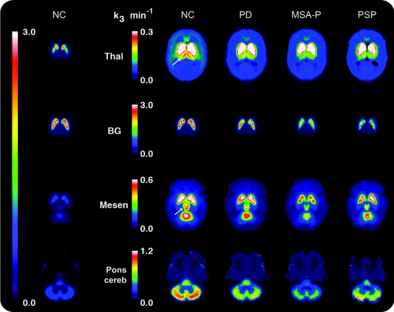
Methods: We used PET with [(11)C]PMP to measure acetylcholinesterase (AChE) activity in multiple cerebral cortical and subcortical regions. We studied 22 normal controls, 12 patients with PD, 13 patients with MSA-P, and 4 patients with PSP.
Results: We found significantly decreased AChE activity in most cerebral cortical regions in PD and MSA-P, and a similar but nonsignificant decrease in PSP. No differences were found between PD and MSA-P. Significantly decreased AChE activity was found in PD in striatum, cerebellum, and thalamus, with a marginally significant decrease in mesencephalon and no change in pons. Significantly greater declines in AChE activity in all subcortical regions were seen in MSA-P and PSP vs in PD. Decreased AChE activity in brainstem and cerebellum of all 3 disorders correlated with disturbances of balance and gait.
Conclusions: Cerebral cortical cholinergic activity is decreased to a similar level in Parkinson disease (PD), parkinsonian syndromes of multiple system atrophy (MSA-P), and progressive supranuclear palsy (PSP) as compared to normal controls. Subcortical cholinergic activity is significantly more decreased in MSA-P and PSP than in PD. The more substantial decrease reflects greater impairment in the pontine cholinergic group, which is important in motor activity, particularly gait. These differences may account for the greater gait disturbances in the early stages of MSA-P and PSP than in PD.
Figures
Comment in
-
Emerging in vivo evidence of subcortical cholinergic dysfunction in Parkinsonian syndromes.Neurology. 2010 May 4;74(18):1406-7. doi: 10.1212/WNL.0b013e3181dfc94e. Neurology. 2010. PMID: 20439841 No abstract available.
References
-
- Davies P, Maloney AJ. Selective loss of central cholinergic neurons in Alzheimer's disease. Lancet 1976;2:1403. - PubMed
-
- Coyle JT, Price DL, DeLong MR. Alzheimer's disease: a disorder of cortical cholinergic innervation. Science 1983;219:1184–1190. - PubMed
-
- Whitehouse PJ, Hedreen JC, White CL 3rd, Price DL. Basal forebrain neurons in the dementia of Parkinson disease. Ann Neurol 1983;13:243–248. - PubMed
-
- Candy JM, Perry RH, Perry EK, et al. Pathological changes in the nucleus of Meynert in Alzheimer's and Parkinson's diseases. J Neurol Sci 1983;59:277–289. - PubMed
-
- Arendt T, Bigl V, Arendt A, Tennstedt A. Loss of neurons in the nucleus basalis of Meynert in Alzheimer's disease, paralysis agitans and Korsakoff's Disease. Acta Neuropathol 1983;61:101–108. - PubMed
Publication types
MeSH terms
Substances
Grants and funding
- R01 AG 19360/AG/NIA NIH HHS/United States
- P01 NS15655/NS/NINDS NIH HHS/United States
- P50 AG08671/AG/NIA NIH HHS/United States
- HL089918/HL/NHLBI NIH HHS/United States
- R01 HL079540/HL/NHLBI NIH HHS/United States
- AG08671/AG/NIA NIH HHS/United States
- UL1RR024986/RR/NCRR NIH HHS/United States
- HL083129/HL/NHLBI NIH HHS/United States
- HD38461/HD/NICHD NIH HHS/United States
- HL087819/HL/NHLBI NIH HHS/United States
- R01 DA016423/DA/NIDA NIH HHS/United States
- P01-CA59827/CA/NCI NIH HHS/United States
- NS015655/NS/NINDS NIH HHS/United States
- UO1 AG024904-01/AG/NIA NIH HHS/United States
- NS044233/NS/NINDS NIH HHS/United States
- HL080941/HL/NHLBI NIH HHS/United States
- P01-CA85878/CA/NCI NIH HHS/United States
- NS42698/NS/NINDS NIH HHS/United States
- NS15655/NS/NINDS NIH HHS/United States
- M01-RR00042/RR/NCRR NIH HHS/United States
- NS02009/NS/NINDS NIH HHS/United States
- U01 AG016976/AG/NIA NIH HHS/United States
- 5 P50 NS15655/NS/NINDS NIH HHS/United States
- 1P01 GM 067189-01A2/GM/NIGMS NIH HHS/United States
- U01-AG16976/AG/NIA NIH HHS/United States
- RR000042/RR/NCRR NIH HHS/United States
- HL03645/HL/NHLBI NIH HHS/United States
- R01 DA022520/DA/NIDA NIH HHS/United States
LinkOut - more resources
Full Text Sources
Medical
Miscellaneous