. 2010 Jun 1;184(11):6479-91.
doi: 10.4049/jimmunol.0903867. Epub 2010 May 3.

Surfactant protein-A as an anti-inflammatory component in the amnion: implications for human pregnancy

Affiliations

Surfactant protein-A as an anti-inflammatory component in the amnion: implications for human pregnancy

Deug-Chan Lee et al. J Immunol. .

Abstract

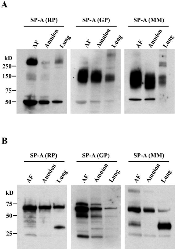
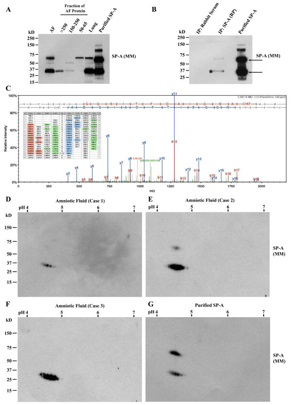
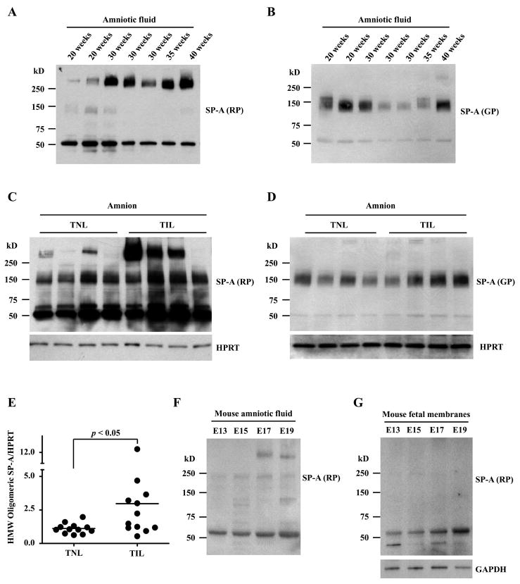
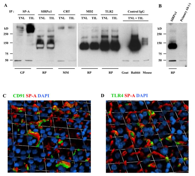
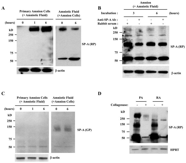
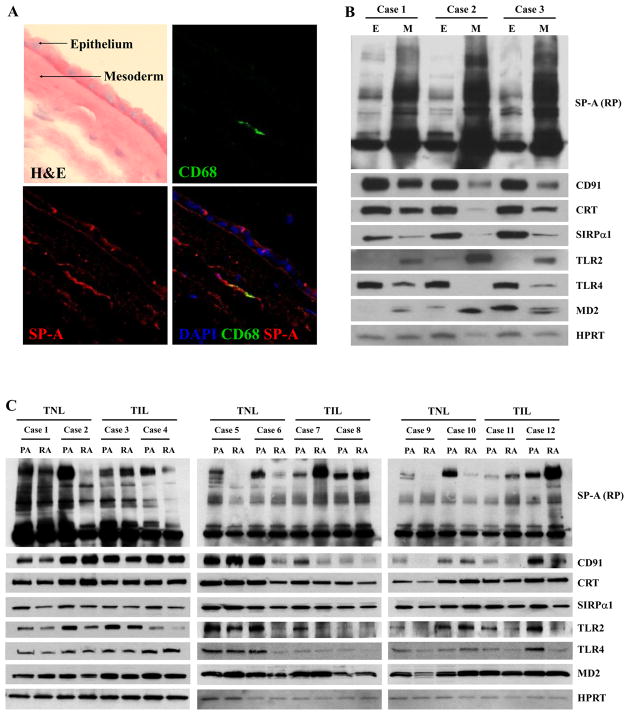
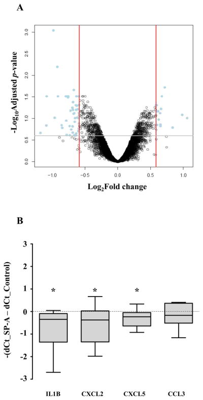
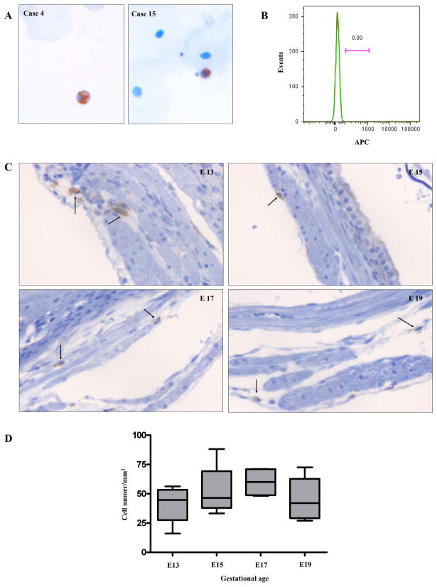
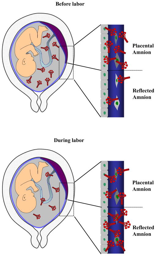
The mechanism of mouse parturition is thought to involve myometrial infiltration by amniotic fluid (AF) macrophages, activated by surfactant protein-A (SP-A). In humans, the concentration of AF SP-A decreases during labor, and no fetal macrophages are found in the myometrium after labor. Therefore, it appears that the mechanisms of labor in mice and humans are different. We investigated a potential role for SP-A in human pregnancy and parturition by examining SP-A expression patterns in AF and amnion. High molecular mass (>250 kDa) oligomeric SP-A was increased in AF with advancing gestation. Interestingly, these oligomers were more abundant in placental amnion before labor at term, while they increased primarily in reflected amnion during labor (p < 0.05). Immunoblotting showed a binding of high molecular mass SP-A in AF to amnion. In C57BL/6 mice, oligomeric SP-A was also readily detected in AF from E15 onwards, but not in amnion. Macrophage density in mice myometrium did not change with advancing gestational age. Microarray analysis of human amnion explants incubated with SP-A revealed a molecular signature of inhibited cytokine-cytokine receptor interaction with downregulation of IL-1beta, CXCL2, and CXCL5 mRNA expression. The findings in this study strongly suggest that SP-A signals amniotic anti-inflammatory response via AF during pregnancy. We propose that an SP-A interaction among AF, placental amnion, and reflected amnion is a unique mechanism for immunoregulation in human pregnancy akin to that established in lung biology. However, AF SP-A and fetal macrophages by themselves do not seem to be exclusive effectors of parturition in humans.


Figures

Figure 1
Figure 1
Immunoreactive patterns of SP-A in human amniotic fluid, amnion, and fetal lung samples to Abs generated from different species (rabbit polyclonal, RP; goat polyclonal, GP; mouse monoclonal, MM) and specificity of immunoreactive bands. A, Two microliters of amniotic fluid and 10 micrograms of total proteins from the amnion and the lung obtained from the same patient were electrophoresed in 4–15% gradient SDS-PAGE under non-reducing conditions, and subsequently electro-blotted. Under non-reducing conditions, the RP anti-SP-A Ab detects HMW (>250 kD) SP-A oligomers and dimers most effectively, whereas GP and MM anti-SP-A Abs effectively recognize MMW (75 kD–250 kD) SP-A oligomers. B, Under reducing conditions, non-reducible SP-A dimers (64 kD) are readily detected by both RP and GP Abs. Monomeric SP-A polypeptides are detected by all Abs, but, most effectively, by the MM Ab.
Figure 2
Figure 2
Characterization of SP-A in the amniotic fluid. A, Amniotic fluid (AF) proteins in slices corresponding to HMW and MMW bands on a non-reducing SDS-PAGE gel were extracted and resolved again by SDS-PAGE under reducing conditions. AF obtained from a twin pregnancy at 29 weeks, and fetal lung tissue from an autopsy case with gestational age of 33 weeks were compared. Immunoblotting with mouse monoclonal anti-SP-A Ab recognizes monomeric SP-A polypeptides in both HMW and MMW fractions, while the 50–65 kD fraction demonstrates largely non-reducible dimers (64 kD). B, Immunoblotting using MM anti-SP-A Ab of HMW SP-A fraction in the same AF (29 weeks) immunoprecipitates with RP anti-SP-A Ab and purified SP-A demonstrates an identical pattern showing monomeric SP-A (32 kD) and non-reducible SP-A dimer (64 kD). C, Mass spectra of monomeric SP-A shown in Figure 2B (*) on LC-MS/MS analysis. LC-MS/MS analysis also confirmed SP-A in monomeric and dimeric SP-A (arrows) in purified SP-A (data not shown). D, E, F, and G, Two-dimensional electrophoresis and SP-A immunoblotting profiles of amniotic fluid samples and purified SP-A. HMW SP-A fraction of AF proteins from three cases (D, 29 weeks; E, 38 weeks; F, 37 weeks). G, Purified SP-A. Electrophoresis was done under reducing conditions followed by immunoblotting with MM anti-SP-A Ab demonstrates the same isomer patterns as that of purified SP-A. However, the upper bands representing non-reducible dimers were hardly detected in all of the AF samples compared with purified SP-A.
Figure 3
Figure 3
Expression patterns of SP-A in the amniotic fluid and the amnions of humans and C57BL/6 mice. A and B, Immunoblotting analysis of amniotic fluid samples obtained across gestational periods shows a clear correlation between gestational age and the HMW oligomeric SP-A expression level detected by the RP anti-SP-A Ab. Two microliters of amniotic fluid samples were electrophoresed in 4–15% gradient SDS-PAGE under a non-reducing condition. C and D, SP-A immunoblotting of the reflected amnion samples shows more prominent HMW (>250 kD) oligomeric SP-A in the cases with labor (TNL: term not in labor, TIL: term in labor). The difference is less prominent for MMW (75 kD-250 kD) SP-A which was detected using the GP anti-SP-A Ab. Ten micrograms of amnion samples were electrophoresed in 4–15% gradient SDS-PAGE under a non-reducing condition. E, Densitometric comparison of HMW SP-A oligomers shows a significant increase of HMW SP-A oligomers in the reflected amnion of TIL cases compared to TNL cases. F, Immunoblotting of C57BL/6 mouse amniotic fluid using the RP anti-SP-A Ab also shows increasing oligomeric SP-A as gestation progresses. HMW SP-A oligomers are readily detectable in E17. Two microliters of amniotic fluid samples were electrophoresed in 4–15% gradient SDS-PAGE under a non-reducing condition. G, In contrast to the findings for the human amnion, mouse fetal membranes do not show oligomeric forms of SP-A. Ten micrograms of fetal membrane samples were electrophoresed in 4–15% gradient SDS-PAGE under a non-reducing condition.
Figure 4
Figure 4
Binding of SP-A to SP-A receptors in the human amnion. A, Human amnion protein lysates (125μg) were immunoprecipitated using specific Abs to SP-A and known SP-A receptors, such as SIRPα, calreticulin (CRT), MD2, and TLR2, and subsequently probed with anti-SP-A Ab. For isotype control experiments to assess specificities of immunoprecipitative reaction, equal amount of proteins were pooled from the same TNL and TIL cases and used. The blot demonstrates co-immunoprecipitation of oligomeric SP-A to SIRPα, CRT, MD2, and TLR2. The species of the primary Ab host used for immunoprecipitation reaction are shown at the bottom of the gel. SP-A immunoblottings were done using a rabbit polyclonal antibody. B, The result of immunoprecipitation in the absence of primary Ab is shown next to the one from SIRPα immunoprecipitation. The same pooled proteins (TNL + TIL) were used. C and D, Immunofluorescent images of the amnion show distinct co-localizations of SP-A with CD91 (C) or TLR4 (D) in the amnion epithelium. Immunofluorescent staining was performed on placental amnion whole mount samples obtained from three TNL cases, and the images were taken from the amnion epithelial layer of a representative case.
Figure 5
Figure 5
Binding of amniotic fluid SP-A oligomers to the human amnion. A, Primary amnion cells incubated with amniotic fluid show an increase in HMW SP-A oligomers in a time-dependent manner (left), while they are being depleted in the conditioned amniotic fluid (right). B, The addition of a neutralizing Ab in the amniotic fluid inhibits HMW SPA binding to the amnion explants. C, The MMW SP-A oligomers are not detected in the amnion cells after incubation with amniotic fluid (left), nor were they changed in the conditioned amniotic fluid (right). D, The SP-A oligomers in the placental amnion (PA) and the reflected amnion (RA) were decreased after treatment with collagenase.
Figure 6
Figure 6
Distribution of SP-A in the human amnion. A, Immunofluorescent staining for SP-A and CD68, a macrophage marker, demonstrates SP-A immunoreactivity in both the amniotic epithelial and mesodermal layers. In the epithelial layer, the signals are detected in both apical portion and basal portion of the cells (original magnification X630). B, Serial immunoblotting for SP-A and its potential receptors in the amnion epithelial layer (E) and the mesodermal layer (M). SP-A is more abundant in the mesodermal layer (p < 0.05). Interestingly, the majority of the potential SP-A receptors was more abundant in the amnion epithelial layer than in the mesodermal layer (p < 0.05), with the exception of TLR2 and MD2, whose expression is clearly higher in the mesodermal layer (p < 0.05). C, Immunoblotting shows more abundant SP-A in the placental amnion (PA) before labor, and SP-A distribution between the placental amnion and the reflected amnion (RA) becomes similar in labor due to an increase in the reflected amnion and a decrease in the placental amnion. The difference in the distribution of HMW SP-A between TNL and TIL cases was significant on densitometric analysis of HMW SP-A oligomers in PA and RA (p < 0.01). The median RA/PA SP-A density ratio was 0.27 (range: 0.13–0.77) in TNL cases, while it was 1.86 (range: 0.4–3.27) in TIL cases. Among the potential receptors screened, the expression of CD91 (p = 0.05) and SIRPα (p = 0.07) tended to be higher in the PA than in the RA. TLR2 (p = 0.14) and TLR4 (p = 0.14) expressions were not significantly different between PA and RA.
Figure 7
Figure 7
Effects of SP-A treatment in the human amnion explants. A, Human reflected amnion explants obtained from TNL patients were treated with SP-A (1 μg/ml) for 12 h. A volcano plot showing the relationship between their fold changes (log2 of, x axis) and FDR-corrected p values (−log10 of, y axis). Positive values on the x-axis indicate genes whose expression increased after SP-A treatment, while negative values indicate genes down-regulated after SP-A treatment. B, Confirmative qRT-PCR data showing changes in the expression of select cytokines and chemokines. IL-1β, CXCL2, and CXCL5 mRNA expressions decreased significantly after treatment with SP-A (p < 0.05 for all comparisons). CCL3 mRNA expression was not significantly different. * p<0.05 by Wilcoxon signed rank test
Figure 8
Figure 8
Macrophages in the human amniotic fluid (A and B) and in the myometrium of C57BL/6 mice (C and D). A, Amniotic fluid cytologic specimens show CD68 immuno-positive macrophages among scattered epithelial cells. Case 4: a case of a patient presenting with intrauterine fetal demise at the gestational age of 21.4 weeks, Case 15: a case of a TNL patient at the gestational age of 38.7 weeks (original magnification X200; hematoxylin was used for counterstaining). B, A histogram from flow cytometric analysis of amniotic fluid cells does not show CD14 positive macrophages. The amniotic fluid was obtained from a TNL patient at the gestational age of 38 weeks. Green: CD14, Red: isotype. C, F4/80 immuno-positive macrophages (arrows) in the C57BL/6 myometrium obtained at various gestational ages: E13, E15, E17, and E19 original magnification X200; hematoxylin was used for counterstaining). D, The density of macrophages in the mouse myometrium did not change significantly across gestation. The numbers of F4/80 positive macrophages were counted in myometrial sections containing the mid-plane of the placenta from four pregnant mice at each gestational age (E13, E15, E17, and E19). Eight high-power fields corresponding to every 45° angle point starting from the mesometrial border in a clockwise direction, covering the entire myometrial circumference, were evaluated in each section. Image-Pro Plus 6.2 software (Media Cybernetics, Inc.) was used for the analysis.
Figure 9
Figure 9
A schematic representation of SP-A dynamics among the amniotic fluid, placental amnion (amnion overlying the placental disc), and the reflected amnion (amnion of the extraplacental chorioamniotic membranes). There is a redistribution of SP-A in the amniotic fluid and the amnion with labor. HMW SP-A oligomers are more abundant in the placental amnion compared to the reflected amnion before labor. With labor at term, increased binding of HMW SP-A oligomers to the reflected amnion results in a decrease of amniotic fluid SP-A. Decreased amniotic fluid SP-A concentration during labor (23) is consistent with this model.

References

    1. Haddad R, Tromp G, Kuivaniemi H, Chaiworapongsa T, Kim YM, Mazor M, Romero R. Human spontaneous labor without histologic chorioamnionitis is characterized by an acute inflammation gene expression signature. Am J Obstet Gynecol. 2006;195:394.e1–24. - PMC - PubMed
    1. Han YM, Romero R, Kim JS, Tarca AL, Kim SK, Draghici S, Kusanovic JP, Gotsch F, Mittal P, Hassan SS, Kim CJ. Region-specific gene expression profiling: novel evidence for biological heterogeneity of the human amnion. Biol Reprod. 2008;79:954–961. - PMC - PubMed
    1. Millar LK, Stollberg J, Debuque L, Bryant-Greenwood G. Fetal membrane distention: determination of the intrauterine surface area and distention of the fetal membranes preterm and at term. Am J Obstet Gynecol. 2000;182:128–134. - PubMed
    1. Mitchell CM, Johnson RF, Giles WB, Zakar T. Prostaglandin H synthase-2 gene regulation in the amnion at labour: histone acetylation and nuclear factor kappa B binding to the promoter in vivo. Mol Hum Reprod. 2008;14:53–59. - PubMed
    1. Romero R, Durum S, Dinarello CA, Oyarzun E, Hobbins JC, Mitchell MD. Interleukin-1 stimulates prostaglandin biosynthesis by human amnion. Prostaglandins. 1989;37:13–22. - PubMed

Publication types

MeSH terms

Substances