

Review
doi: 10.1083/jcb.201002027.
The immunological synapse: a focal point for endocytosis and exocytosis
Affiliations
- PMID: 20439993
- PMCID: PMC2867296
- DOI: 10.1083/jcb.201002027
Item in Clipboard
Review
The immunological synapse: a focal point for endocytosis and exocytosis
J Cell Biol.
.
Abstract
There are many different cells in the immune system. To mount an effective immune response, they need to communicate with each other. One way in which this is done is by the formation of immunological synapses between cells. Recent developments show that the immune synapse serves as a focal point for exocytosis and endocytosis, directed by centrosomal docking at the plasma membrane. In this respect, formation of the immunological synapse bears striking similarities to cilia formation and cytokinesis. These intriguing observations suggest that the centrosome may play a conserved role in designating a specialized area of membrane for localized endocytosis and exocytosis.
Figures
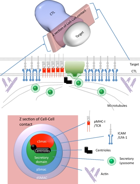
The immunological synapse. Cartoon summary of the organization of receptors showing the relative positions of the cSMAC, pSMAC, dSMAC, centrioles, actin and microtubule cytoskeletons, and secretory lysosomes at the immunological synapse in cross section and across the area of cell contact.
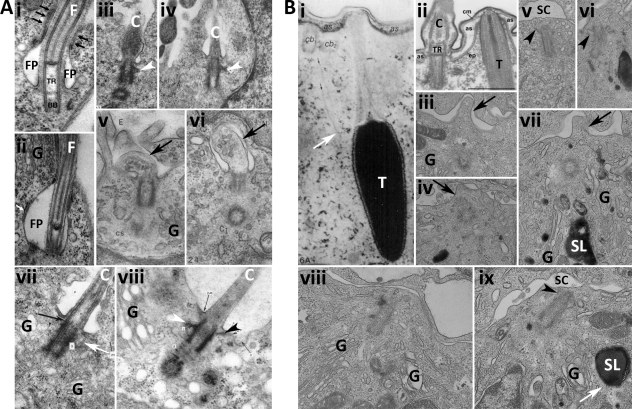
Electron micrographs comparing flagella, primary cilia, trichocyst secretion, and the immunological synapse. (A) EM images of flagella (i and ii) and primary cilia (C; iii–viii) showing basal bodies (BB), flagella (F), flagella pocket (FP), transition region (TR), Golgi (G), distal (black arrowheads) and subdistal (white arrowheads) appendages of the mother centriole, membrane protrusions formed during ciliogenesis (black arrows in v and vi), and microtubules linking the centriole to the membrane (white arrow in vii). An area of tight contact between the flagella pocket and flagella (black arrows in i) and a flat compartment close to the flagella pocket (white arrow in ii) are also shown. Basal body fibers and cilia fibers are contiguous (black arrow in vii). Images were reproduced from Absalon et al. (2008; i and ii; with permission from the Journal of Cell Science), Archer and Wheatley (1971; iii; with permission from Wiley-Blackwell), Wheatley (1971; iv and vii; with permission from Wiley-Blackwell), Sorokin (1962; v and vi), and Wheatley (1967; viii; with permission from Wiley-Blackwell). (B) EM images of trichocyst (T) secretion from paramecia (i and ii) showing cilia (C), ciliary basal bodies (cb), alveolar sacs (as), cell membrane (cm), epiplasm (ep), and transition region (TR) and immune synapses formed between CTLs (lower cell) and targets (upper cell; iii–ix) showing Golgi (G), secretory lysosomes (SL), synaptic cleft (SC), membrane bulges (black arrows), mother centriole appendages (black arrowheads), and microtubules associated with microtubules (white arrows). Images were reproduced from Glas-Albrecht et al. (1991; i; with permission from the Journal of Cell Science), Plattner (2002; ii; with permission from Wiley-VCH Verlag GmbH & Co. KGaA and the author). CTL images are from J.C. Stinchcombe.
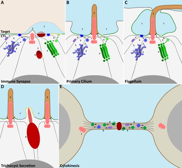
A common theme in the architecture of diverse cellular structures. Cartoon illustrating comparison of the organization of the immune synapse (A), primary cilium (B), flagella pocket (C), point of trichocyst secretion in paramecia (D), and site of cytokinesis in dividing cells (E), indicating that the site of centrosome polarization is a focal point for exocytosis and endocytosis in different cellular systems. Similarities between some or all of these examples include (1) polarization of the centrosome (pink) to the plasma membrane; (2) formation of membrane protrusions (brown) opposite the point of centrosome docking; polarized movement of (3) the microtubule cytoskeleton and (4) the Golgi complex (green) and endocytic recycling compartments (mauve) away from the nucleus (gray) toward the site of centrosome docking at the plasma membrane; (5) redirection of the biosynthetic (green), endocytic (blue), and recycling (purple) pathways to the plasma membrane; (6) focused polarization of regulated secretory organelles (e.g., trichocysts [crimson] in paramecia and secretory lysosomes [claret] with secretory cores [crimson] in CTLs) to specialized secretory domains at the plasma membrane (orange) by minus end–directed transport along microtubules; and (7) formation of specialized enclosed (e.g., secretory clefts [CTLs] and flagella pockets) or semienclosed (cilia and trichocyst) extracellular spaces to which endocytosis and exocytosis are focused.
References
-
- Absalon S., Blisnick T., Bonhivers M., Kohl L., Cayet N., Toutirais G., Buisson J., Robinson D., Bastin P. 2008. Flagellum elongation is required for correct structure, orientation and function of the flagellar pocket in Trypanosoma brucei. J. Cell Sci. 121:3704–3716 10.1242/jcs.035626 - DOI - PubMed
Publication types
MeSH terms
Grants and funding
LinkOut - more resources
Full Text Sources
