. 2010 May 3;189(3):527-39.
doi: 10.1083/jcb.200912125.

Lack of Apobec2-related proteins causes a dystrophic muscle phenotype in zebrafish embryos

Affiliations

Lack of Apobec2-related proteins causes a dystrophic muscle phenotype in zebrafish embryos

Christelle Etard et al. J Cell Biol. .

Abstract

The chaperones Unc45b and Hsp90a are essential for folding of myosin in organisms ranging from worms to humans. We show here that zebrafish Unc45b, but not Hsp90a, binds to the putative cytidine deaminase Apobec2 (Apo2) in an interaction that requires the Unc45/Cro1p/She4p-related (UCS) and central domains of Unc45b. Morpholino oligonucleotide-mediated knockdown of the two related proteins Apo2a and Apo2b causes a dystrophic phenotype in the zebrafish skeletal musculature and impairs heart function. These phenotypic traits are shared with mutants of unc45b, but not with hsp90a mutants. Apo2a and -2b act nonredundantly and bind to each other in vitro, which suggests a heteromeric functional complex. Our results demonstrate that Unc45b and Apo2 proteins act in a Hsp90a-independent pathway that is required for integrity of the myosepta and myofiber attachment. Because the only known function of Unc45b is that of a chaperone, Apo2 proteins may be clients of Unc45b but other yet unidentified processes cannot be excluded.


Figures

Figure 1.
Figure 1.
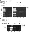
Apo2a interacts with Unc45b but not Hsp90a in pull-down assays. (A) Schematic representation of the full-length Unc45b protein fused to GFP. Unc45b is composed of three domains: the TPR (amino acids 1–119), the central (amino acids 120–509), and the UCS (amino acid 910–935) domains. The intact Unc45b protein and its subdomains as well as Hsp90a were fused at their C terminus to GFP and used as input protein in the pull-down assays as indicated. (B) In vitro synthesized chimeric proteins visualized by Western blotting with anti-GFP antibody. (C) Full-length Unc45b-GFP fusion protein (lane 1) and deletion variants of Unc45b fused to GFP (TPR-deleted Unc45b [Unc45-ΔTPR] lane 2; UCS-deleted Unc45b [Unc45b-ΔUCS], lane 3; or single Unc45b domains fused to GFP [Unc45b-Central], lane 4; Unc45b-TPR, lane 5; Unc45b-UCS, lane 6) were pulled down with the GST-Apo2a fusion protein. With the exception of Unc45b-TPR (lane 5), all other chimeras were pulled down by GST-Apo2a fusion proteins (lanes 1–4 and 6). (D) Western blot of protein input of Hsp90a-GFP fusion (lane 11). Unc45b-UCS (lane 8), Unc45b-TPR (lane 9), and GFP (lane 10) were used as positive and negative controls, respectively, to test for an interaction of GST-Apo2a with Hsp90a-GFP. Note that relative to protein inputs in binding assays (C and E), 10-fold less protein was loaded onto the control gels shown in B and D. White lines indicate that intervening lanes have been spliced out. (E) Although Unc45b-UCS interacted with Apo2a (lane 8), neither the Unc45b-TPR fusion (lane 9), GFP alone (lane 10), nor Hsp90a-GFP (lane 11) were pulled down by GST-Apo2a. Thus, the interaction of Apo2a with Unc45b is specific. Lane 7 and 12 show protein markers. White boxes indicate 95 kD.
Figure 2.
Figure 2.
Apo2a is expressed in the heart and in the skeletal musculature. (A–C) In situ hybridization with an apo2a anti-sense probe to 18 somites (A) and 24-hpf (B and C) embryos showing apo2a mRNA expression in cardiac (h) and skeletal muscles (s). (D–F) At 48 (D and E) and 60 hpf (F), the expression was found in the somitic muscle (s; E and F), hypaxial muscles (hy; F), the pectoral fin muscles (pf; E), and cranial muscles (adductor mandibulae [am], adductor hyomandibulae [ah], sternohyoideus [sh], constrictor hyoideus ventralis [chv], and inferior oblique [io]; D and E). The orientation of embryos in the images is anterior to the left and are as follows: (A) dorso-lateral view; (B, C, and E) lateral view, dorsal up; (D) ventral view; and (F) dorsal view. Bars, 200 µm.
Figure 3.
Figure 3.
Apo2a morphants have defects in the skeletal musculature and the heart. (A) Position of the morpholinos Mo(ATG)-apo2a (ATG) and Mo(UTR)-apo2a (UTR) on the apo2a mRNA. (B–D) Mo(ATG)-apo2a (B and D) and control Mo(ATG)-apo2a-cont (harboring five mismatches relative to the Mo(ATG)-apo2a sequence; C)-injected embryos. The morphants exhibit a curved body axis (B) and show reduced birefringence (D) compared with controls (C). Note that the Mo(ATG)-apo2a–injected embryos (B and D) were coinjected with the Mo-p53 to suppress unspecific effects (see K–M for additional controls for Mo-p53 injection). (E–G) Mo(UTR)-apo2a (E and G)- and control Mo(ATG)-apo2a-cont (F)-injected embryos. The morphants exhibit a curved body axis (E) and show reduced birefringence (G) compared with controls (F). (H–J) Apo2a-GFP expression (H) in embryos injected with an apo2a-gfp encoding plasmid. Co-injection of Mo(ATG)-apo2a with the apo2a-gfp plasmid abolishes Apo2a-GFP expression (I), whereas coinjection of the apo2a-gfp plasmid with the five-mismatch Mo(ATG)-apo2a-cont does not abolish Apo2a-GFP expression (J). (K–M) Uninjected (K), Mo(ATG)-apo2a-cont– and Mo-p53–coinjected (L), and Mo(ATG)-apo2a– and Mo-p53–coinjected embryos (M). Injection of Mo(ATG)-apo2a morpholino reduces birefringence (M), whereas neither the five-mismatch Mo(ATG)-apo2a-cont nor the Mo-p53 had an effect on the birefringence of the musculature (L). (N) Mo(ATG)-apo2a and Mo(UTR)-apo2a morphants have a decreased heart rate (the graph shows heart beats per minute, n = 20 embryos). Black bars represent standard deviation. The datasets were pairwise subjected to the Student’s t test. The difference between the heart rate of Mo(ATG)-apo2a versus Mo(UTR)-apo2a was not significant (P = 0.220), whereas the difference between the heart rate of Mo(ATG)-apo2a versus control or Mo(UTR)-apo2a versus control was highly significant (P = 0.002 and P = 0.003, respectively). (O) Pericardial edema (arrow) in a Mo(ATG)-apo2a– and Mo-p53–coinjected embryo. Embryos shown are 72 hpf, except F–H, which show 24-hpf embryos. Pigmentation of the embryo shown in E was suppressed by 1-phenyl 2-thiourea (PTU) treatment (Karlsson et al., 2001). Bars, 80 µm.
Figure 4.
Figure 4.
Apo2a morphants have defective myosepta and form intersomitic giant myofibers. (A–C) DIC images of Mo(ATG)-apo2a–injected embryos. Mo(ATG)-apo2a morphants (Mo) exhibit cell-free spaces in the somites (A and B, stars), disrupted myosepta (B and C, arrows), and giant myofibers that span two somites (C, arrowhead). (D–F) Immunohistochemistry with antibodies directed against myoseptal proteins reveals defects in the myosepta of apo2a morphants: α-Dystroglycan (DG) staining of control (cont; D) and Mo(ATG)-apo2a–injected (Mo; E and F) embryos (white arrows, disrupted myosepta; green arrows, split myosepta). (G and H) Laminin staining reveals abnormal myosepta in Mo(ATG)-apo2a morphants (H) compared with wild type (G). Green arrows, split myosepta. (I–K) Double immunohistochemistry with anti–α-dystroglycan (DG) and the anti-slow muscle myosin antibody F59 reveals giant slow muscle myofibers (J, arrowhead) crossing the somite boundary (J, arrow). (I) Control embryos never show these intersomitic myofibers. Detachment of myofibers from the myosepta (K, arrow) generates cell-free spaces (K, star). (L and M) Sarcomeric myosin stained with A4.1025 in Mo(ATG)-apo2a morphants shows detached slow fibers (M, yellow arrows) overlaying fast fibers and generating cell-free areas inside the muscle tissue (star). Most of the fast fibers are still anchored to the myosepta. A few fast fibers have also detached (M, blue arrow). (L) Control. All embryos injected with Mo(ATG)-apo2a were coinjected with Mo-p53. Embryos are 72 h old. The anterior is shown to the left, dorsal side up. Bars, 25 µm.
Figure 5.
Figure 5.
Apo2b interacts with Unc45b. (A) Schematic diagram of the Unc45b-GFP full-length and deletion proteins used in pull-down assays. (B and C) Western blot of in vitro translated input (B) and Unc45b-GFP proteins pulled down with GST-Apo2b (C). Fusion proteins were detected with an anti-GFP antibody. With the exception of Unc45b-TPR (lane 5), all the other fusions proteins were pulled down by Apo2b-GST (lanes 1–4 and 6), which indicates that both the central as well as the UCS domain can mediate the interaction with Apo2b. Lane 7 shows size markers. Note that relative to protein inputs in binding assays (B), 10-fold less protein was loaded onto the control gels shown in C. White boxes indicate 95 kD. (D and E) Control (D) and Mo(ATG)-apo2b (E)-injected embryos. Injection of the morpholino complementary to sequences surrounding the ATG of apo2b mRNA reduces the birefringence of the skeletal muscle. (F and G) Control (F) and Mo(UTR)-apo2b (G)-injected embryos. The morpholino complementary to the 5′ UTR of the apo2b mRNA reduces the birefringence of the skeletal muscle in a similar manner to the morpholino directed against the ATG of apo2b mRNA. The striking similarity of this phenotype to apo2a morphants suggests that the two Apo2 proteins do not act redundantly. (H and I) Mo(ATG)-apo2b morphants (I) but not controls (H) observed with DIC optics have cell-free spaces in the somites (I, stars). (J and K) Double immunohistochemistry with a slow muscle myosin antibody F59 and/or an α-dystroglycan (DG; J and K, respectively) antibody reveals extra-long intersomitic myofibers spanning two somites (K, arrowhead), detached myofibers, and defective myosepta in the morphants (K; white arrow, disrupted myosepta; yellow arrow, detached fibers). (I) Control. Bars: (D, E, and H–K) 30 µm; (F) 80 µm; (G) 100 µm.
Figure 6.
Figure 6.
Apo2a and Apo2b can form homo- and heteromers. (A) GST-Apo2a and GST-Apo2b fusion proteins can pull down Apo2b-GFP proteins. Pulled-down proteins were detected by Western blotting with an anti-GFP antibody. Lanes 1 and 4: protein size markers (white boxes indicate 95 kD); lanes 2 and 5: GFP-only controls; lane 3: pull-down of Apo2b-GFP by GST-Apo2a; lane 6: pull-down of Apo2b-GFP by GST-Apo2b; lane 7: Apo2b-GFP protein input. (B) GST-Apo2a and GST-Apo2b can pull down MYC-Apo2a fusion proteins as revealed by Western blotting with the anti-MYC epitope 9E10 antibody. Lane 1: pull-down of Apo2a-MYC with GST-Apo2a; lane 2: protein size markers (white boxes indicate 72 kD); lane 3: input of MYC-Apo2a protein; lane 4: GST-only control; lane 5: pull-down of MYC-Apo2a by GST-Apo2b.
Figure 7.
Figure 7.
unc45b mutants also exhibit cell-free spaces in the somites and disruption of the myosepta like the apo2a morphants. (A–C) α-Actin–GFP transgenic wild-type (A), unc45b (B), and hsp90a (C) mutant embryos. unc45b mutant embryos exhibit cell-free spaces in the somitic musculature (B, stars) as seen in apo2a morphants. In contrast, wild-type siblings (A) and hsp90a (C) mutants do not show these gaps. (D–F) Cell-free spaces are also visible in unlabeled unc45b mutants using DIC optics (E, white stars). In contrast, wild-type siblings (D) and hsp90a mutants (F) do not exhibit these cell-free spaces in the somites. Broken lines indicate myoseptal boundaries. (G) Merge of DIC and α-actin–GFP fluorescence frames shows detachment of myofibrils (arrows) and cell-free spaces (stars) in unc45b mutants, which is seen in apo2a morphants but never seen in wild-type or morphant control embryos. (H and I) Immunohistochemistry with a β-dystroglycan (DG) antibody shows disruption of myosepta in unc45b mutants (I), whereas wild-type siblings (H) have normal chevron-shaped myosepta. (J–L) Unc45b-GFP is localized at the Z line (J, white arrow) as well as at the myosepta (blue arrow), whereas embryos expressing CapZa1-GFP show localization at the Z line exclusively (L, arrow). Immunohistochemistry with an antibody against endogenous Unc45b reveals that endogenous Unc45b is localized at the myosepta (K and K′, blue arrows) in addition to the previously shown Z line staining (K′, white arrow). Bars: (A–I and K) 40 µm; (J and L) 2 µm; (K′): 4 µm.
Figure 8.
Figure 8.
Localization of Apo2a and Apo2b fusion proteins. (A–C) Mosaic expression of Apo2a-mOrange1 fusion proteins in individual cells of the skeletal muscles. Cells expressing Apo2a-mOrange1 at low levels show enrichment of the fusion proteins at the myoseptal boundary (A and B, arrowheads). Low levels and more diffuse staining are present in the cytoplasm and over the myofibril. Enrichment of Apo2a-mOrange1 at the myoseptal boundary overlaps with Unc45b-TFP (C, arrowhead). (D–F) Mosaic expression of Apo2b-mOrange1 fusion proteins in individual cells of the skeletal muscles. Diffuse staining in the cytoplasm and in a striated manner at the Z line of the myofibrils was noted for this fusion protein. Apo2b–mOrange is colocalized with Unc45b over the Z line and also at the myoseptal boundary (D and F, arrowhead) in cells coexpressing Unc45b-TFP fusion protein (C). (G–I) Expression of Apo2a-mOrange1 at high levels results in aggregation of the protein in the cytoplasm (G). This results in loss of striation in highly expressing cells (H and I, arrows; overlay of G and H). (J and K) Lack of Unc45b protein leads to aggregation of Apo2a-mOrange1 (J) and Apo2b-mOrange1 fusion proteins. Bars: (A, C, D, and F) 16 µm; (B, E, J, and K) 4 µm; (G, H, and I) 10 µm.

Similar articles

Cited by

References

    1. Ai H.W., Henderson J.N., Remington S.J., Campbell R.E. 2006. Directed evolution of a monomeric, bright and photostable version of Clavularia cyan fluorescent protein: structural characterization and applications in fluorescence imaging. Biochem. J. 400:531–540 10.1042/BJ20060874 - DOI - PMC - PubMed
    1. Amali A.A., Lin C.J., Chen Y.H., Wang W.L., Gong H.Y., Rekha R.D., Lu J.K., Chen T.T., Wu J.L. 2008. Overexpression of Myostatin2 in zebrafish reduces the expression of dystrophin associated protein complex (DAPC) which leads to muscle dystrophy. J. Biomed. Sci. 15:595–604 10.1007/s11373-008-9250-2 - DOI - PubMed
    1. Anant S., Mukhopadhyay D., Sankaranand V., Kennedy S., Henderson J.O., Davidson N.O. 2001. ARCD-1, an apobec-1-related cytidine deaminase, exerts a dominant negative effect on C to U RNA editing. Am. J. Physiol. Cell Physiol. 281:C1904–C1916 - PubMed
    1. Anderson M.J., Pham V.N., Vogel A.M., Weinstein B.M., Roman B.L. 2008. Loss of unc45a precipitates arteriovenous shunting in the aortic arches. Dev. Biol. 318:258–267 10.1016/j.ydbio.2008.03.022 - DOI - PMC - PubMed
    1. Barral J.M., Bauer C.C., Ortiz I., Epstein H.F. 1998. Unc-45 mutations in Caenorhabditis elegans implicate a CRO1/She4p-like domain in myosin assembly. J. Cell Biol. 143:1215–1225 10.1083/jcb.143.5.1215 - DOI - PMC - PubMed

Publication types

MeSH terms