. 2010 Jun;120(6):2171-83.
doi: 10.1172/JCI35846. Epub 2010 May 3.

Hypoxia-inducible factor-1alpha regulates beta cell function in mouse and human islets

Affiliations

Hypoxia-inducible factor-1alpha regulates beta cell function in mouse and human islets

Kim Cheng et al. J Clin Invest. 2010 Jun.

Abstract

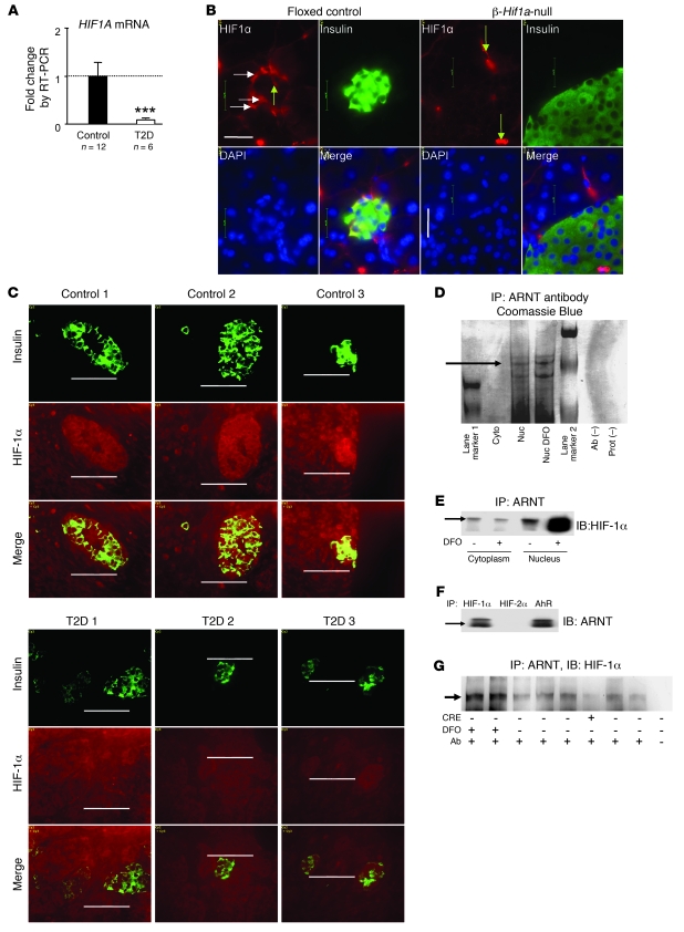
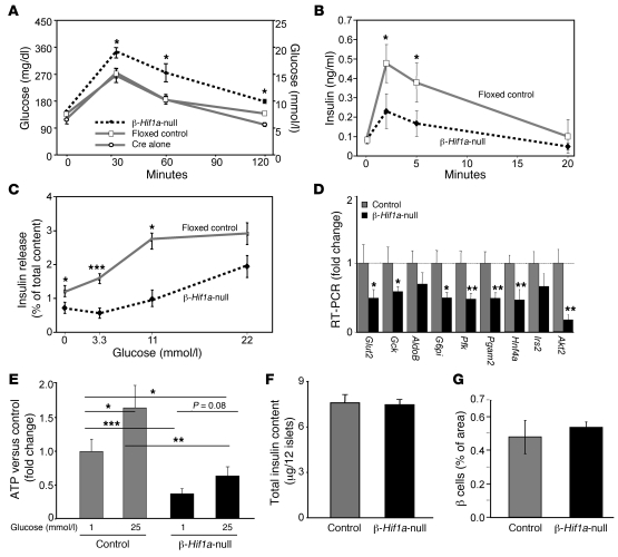
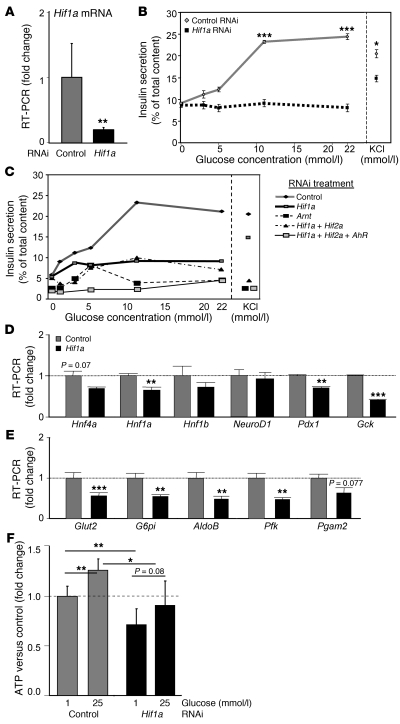
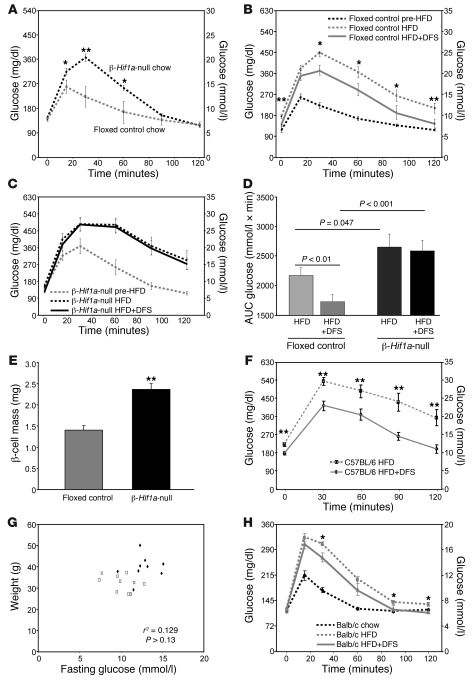
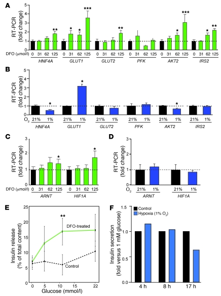
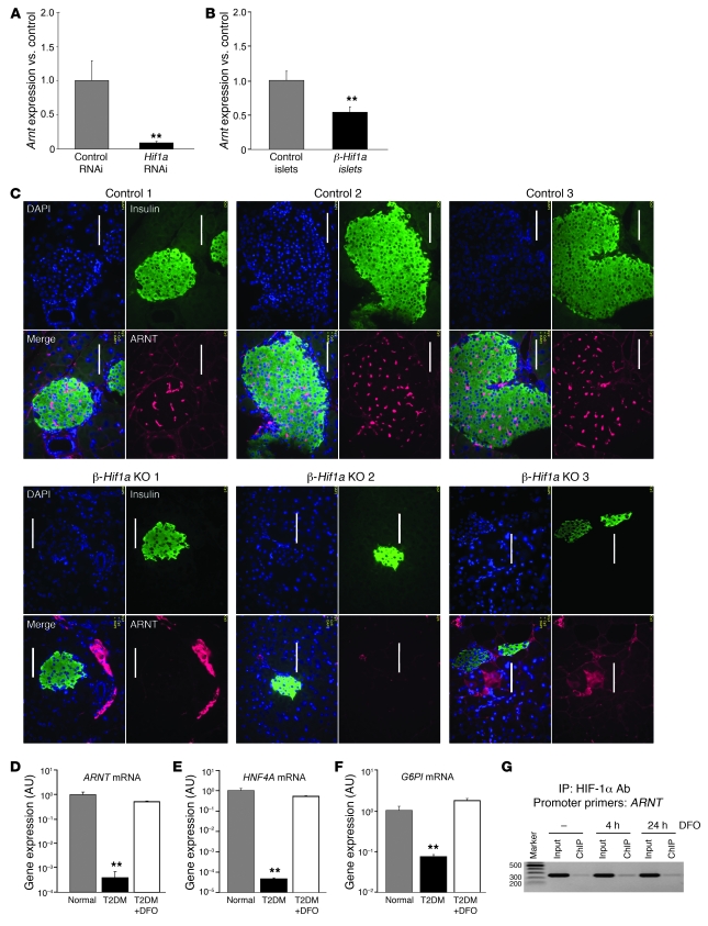
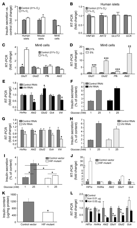
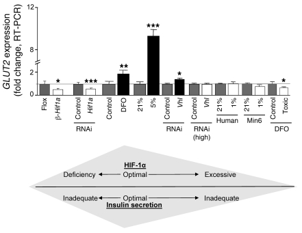
Hypoxia-inducible factor-1alpha (HIF-1alpha) is a transcription factor that regulates cellular stress responses. While the levels of HIF-1alpha protein are tightly regulated, recent studies suggest that it can be active under normoxic conditions. We hypothesized that HIF-1alpha is required for normal beta cell function and reserve and that dysregulation may contribute to the pathogenesis of type 2 diabetes (T2D). Here we show that HIF-1alpha protein is present at low levels in mouse and human normoxic beta cells and islets. Decreased levels of HIF-1alpha impaired glucose-stimulated ATP generation and beta cell function. C57BL/6 mice with beta cell-specific Hif1a disruption (referred to herein as beta-Hif1a-null mice) exhibited glucose intolerance, beta cell dysfunction, and developed severe glucose intolerance on a high-fat diet. Increasing HIF-1alpha levels by inhibiting its degradation through iron chelation markedly improved insulin secretion and glucose tolerance in control mice fed a high-fat diet but not in beta-Hif1a-null mice. Increasing HIF-1alpha levels markedly increased expression of ARNT and other genes in human T2D islets and improved their function. Further analysis indicated that HIF-1alpha was bound to the Arnt promoter in a mouse beta cell line, suggesting direct regulation. Taken together, these findings suggest an important role for HIF-1alpha in beta cell reserve and regulation of ARNT expression and demonstrate that HIF-1alpha is a potential therapeutic target for the beta cell dysfunction of T2D.


Figures

Figure 1
Figure 1. HIF-1α is present in normoxic β cells, associates with ARNT, and is decreased in T2D.
(A) HIF1A mRNA was decreased in islets of people with T2D (n = 6) compared with people with normal glucose tolerance (n = 12). ***P < 0.001. (B) HIF-1α protein was present in β cells in floxed control mice (horizontal arrows) but was decreased in β-Hif1a-null mice. In both genotypes, HIF-1α was present in blood vessels (vertical arrows). Scale bar: 20 μm. (C) HIF-1α protein was higher than background in people with normal glucose tolerance (top panels) but was decreased in T2D pancreata (bottom panels). Scale bar: 50 μm. (D) HIF-1α protein (arrow) was associated with ARNT by affinity purification. Cyto, cytoplasm; Nuc, nucleus; Prot, protein. (E) HIF-1α protein associated with ARNT in the basal state in Min6 cells, and nuclear HIF-1α increased with DFO. (F) ARNT protein associated with HIF-1α by coimmunoprecipitation. (G) HIF-1α protein was increased by DFO treatment of isolated mouse islets and was decreased in islets from a β-Hif1a-null mouse (Cre+).
Figure 2
Figure 2. β cell deletion of Hif1a in mice causes glucose intolerance, impaired gene expression, ATP generation, and insulin secretion.
(A) β-Hif1a-null mice were glucose intolerant compared with either floxed controls or RIP-Cre alone mice. n = 15, 15, and 12, respectively. (B) GSIS was decreased in β-Hif1a-null mice. (C) GSIS was decreased in isolated β-Hif1a-null islets. (D) Expression of several genes was decreased in β-Hif1a-null islets. (E) ATP concentrations were significantly decreased in β-Hif1a-null islets at both basal and high glucose levels. (F) Insulin content did not differ between floxed control and β-Hif1a-null islets. (G) β cell mass did not differ between groups. *P < 0.05, **P < 0.01, and ***P < 0.001.
Figure 3
Figure 3. Decreasing Hif1a by RNAi-impaired β cell function, gene expression, and ATP generation.
(A) RNAi decreased Hif1a mRNA. (B) Hif1a RNAi decreased GSIS in Min6 cells and caused a small decrease in KCl-stimulated insulin release. (C) Combination RNAi treatment caused slightly more severe impairment in insulin release. (D) Hif1a RNAi decreased expression of genes from the MODY family and (E) glucose-uptake and glycolysis genes. (F) Hif1a RNAi decreased basal and glucose-stimulated ATP concentrations. *P < 0.05, **P < 0.01, and ***P < 0.001.
Figure 4
Figure 4. Lack of β cell HIF-1α leads to severe deterioration in glucose tolerance on a HFD and increasing HIF-1α levels with DFS improves glucose tolerance on a HFD.
(A) β-Hif1a-null mice (n = 10) had worse glucose tolerance than floxed control littermates (n = 17). (B) On HFD, glucose tolerance deteriorated in floxed controls and improved following DFS. (C) On HFD, glucose tolerance deteriorated markedly in β-Hif1a-null mice, and there was no improvement with DFS. (D) Glucose tolerance AUC for mice at completion of the HFD and HFD plus DFS stages. (E) β cell mass was increased in β-Hif1a-null mice at study completion. (F) Glucose tolerance was significantly better in C57BL/6 mice receiving HFD plus DFS versus mice receiving HFD alone (n = 10 per group). (G) Weight and fasting glucose were not significantly correlated in the mice. Rectangles indicate mice receiving HFD plus DFS, and triangles indicate mice receiving HFD alone. (H) Balb/c mice had deterioration in glucose tolerance on HFD (dotted line) compared with chow (dashed line). Their glucose tolerance improved significantly on HFD plus DFS (n = 12). *P < 0.05 and **P < 0.01.
Figure 5
Figure 5. DFO improves gene expression and insulin secretion from human islets.
(A) DFO increased expression of several genes in isolated human islets. (B) Hypoxic culture increased GLUT1 expression but decreased that of HNF4A and AKT2. (C) DFO increased expression of ARNT and HIF1A. (D) Hypoxia did not alter ARNT or HIF1A expression. (E) DFO increased insulin secretion in isolated human islets. (F) GSIS was not improved in hypoxic islets and declined with longer exposure. *P < 0.05, **P < 0.01, and ***P < 0.001.
Figure 6
Figure 6. HIF-1α regulates expression of ARNT and downstream genes.
(A) Hif1a RNAi in Min6 cells decreased Arnt expression. (B) β-Hif1a-null mice had decreased Arnt mRNA compared with floxed controls. (C) ARNT protein was decreased in β-Hif1a-null islets versus floxed controls. Scale bar: 50 μm. (D) ARNT mRNA was decreased in islets from people with T2D. DFO increased ARNT expression to levels that did not differ significantly from normal. (E) HNF4A mRNA was decreased in islets from people with T2D and was increased by DFO. (F) G6PI expression was decreased in islets from people with T2D and was increased by DFO. (G) HIF-1α associated with the proximal Arnt promoter by ChIP. **P < 0.01.
Figure 7
Figure 7. The differing effects on gene expression and insulin secretion with increasing HIF-1α levels using hypoxia or VHL RNAi.
(A) Insulin secretion was not improved in human islets, mouse islets, or Min6 cells cultured under hypoxic conditions. (B) One percent oxygen did not increase GLUT2 expression in human islets. (C) One percent oxygen did not increase Glut2 expression in Min6 cells. (D) Moderate hypoxia (5% oxygen) increased Glut2 expression. (E) Modest Vhl knockdown (35%) increased expression of Glut2 and was associated with a nonsignificant increase in insulin release (F). (G) High-dose Vhl knockdown achieved a 55% decrease in Vhl and did not increase Glut2 expression. (H) High-dose Vhl RNAi lowered insulin secretion nonsignificantly. (I) Transfection with proline-to-alanine mutant HIF-1α significantly impaired insulin secretion. (J) Hif1a expression was increased more than 29-fold and was accompanied by increased Glut1 expression and decreased Gck. (K) Total insulin content was decreased in the proline mutant HIF–overexpressing cells. (L) Ferric citrate treatment significantly decreased Hif1a expression and was accompanied by decreased expression of Hnf4a, Akt2, Glut1, and Glut2. *P < 0.05, **P < 0.01, and ***P < 0.001.
Figure 8
Figure 8. Modest increases in HIF-1α improve insulin secretion.
Changes in HIF-1α, which were associated with decreased GLUT2, were associated with impaired insulin secretion. *P < 0.05, **P < 0.01, and ***P < 0.001.

Similar articles

Cited by

References

    1. Hofer T, Wenger H, Gassmann M. Oxygen sensing, HIF-1α stabilization and potential therapeutic strategies. Pflugers Arch. 2002;443(4):503–507. doi: 10.1007/s00424-001-0759-8. - DOI - PubMed
    1. Semenza GL. Regulation of mammalian O2 homeostasis by hypoxia-inducible factor 1. Annu Rev Cell Dev Biol. 1999;15:551–578. doi: 10.1146/annurev.cellbio.15.1.551. - DOI - PubMed
    1. Kotch LE, Iyer NV, Laughner E, Semenza GL. Defective vascularization of HIF-1α-null embryos is not associated with VEGF deficiency but with mesenchymal cell death. Dev Biol. 1999;209(2):254–267. doi: 10.1006/dbio.1999.9253. - DOI - PubMed
    1. Iyer NV, et al. Cellular and developmental control of O2 homeostasis by hypoxia inducible factor 1α. . Genes Dev. 1998;12(2):149–162. doi: 10.1101/gad.12.2.149. - DOI - PMC - PubMed
    1. Cowden Dahl KD, et al. Hypoxia-inducible factors 1{alpha} and 2{alpha} regulate trophoblast differentiation. Mol Cell Biol. 2005;25(23):10479–10491. doi: 10.1128/MCB.25.23.10479-10491.2005. - DOI - PMC - PubMed

Publication types