

Review
doi: 10.1172/JCI41911.
Epub 2010 May 3.
Trafficking of immune cells in the central nervous system
Affiliations
- PMID: 20440079
- PMCID: PMC2860945
- DOI: 10.1172/JCI41911
Item in Clipboard
Review
Trafficking of immune cells in the central nervous system
J Clin Invest.
2010 May.
Abstract
The CNS is an immune-privileged environment, yet the local control of multiple pathogens is dependent on the ability of immune cells to access and operate within this site. However, inflammation of the distinct anatomical sites (i.e., meninges, cerebrospinal fluid, and parenchyma) associated with the CNS can also be deleterious. Therefore, control of lymphocyte entry and migration within the brain is vital to regulate protective and pathological responses. In this review, several recent advances are highlighted that provide new insights into the processes that regulate leukocyte access to, and movement within, the brain.
Figures
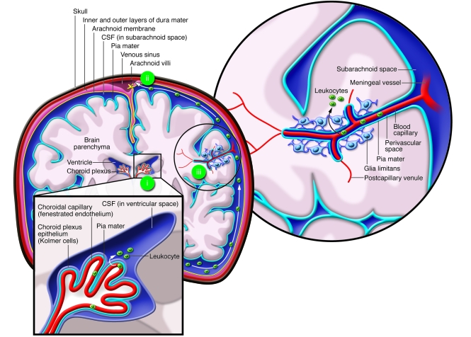
Beneath the skull lie three membranes that enclose the parenchyma of the brain:
the dura mater, the arachnoid membrane, and the pia mater. The latter two enclose
the subarachnoid space. (i) Leukocytes can enter across the choroid plexus, where
CSF is produced by the choroid plexus epithelium in the ventricles. CSF containing
leukocytes then enters the subarachnoid space, circulates around the brain, and
(ii) exits via the venous sinus to be resorbed by the blood via the arachnoid
villi. (iii) Blood supply to the brain enters in the subarachnoid space over the
pia mater, generating the perivascular space (or Virchow-Robin space). Main
arterial branches divide into capillaries, which terminate deep within the brain,
supplying the parenchyma with blood. Leukocytes can potentially enter from the
blood (iii), which requires them to cross the tightly regulated vascular
endothelium (i.e., the BBB: the glia limitans, the subarachnoid space, and the pia
mater). Cells can adhere to the endothelium and arrest at any point during this
process.
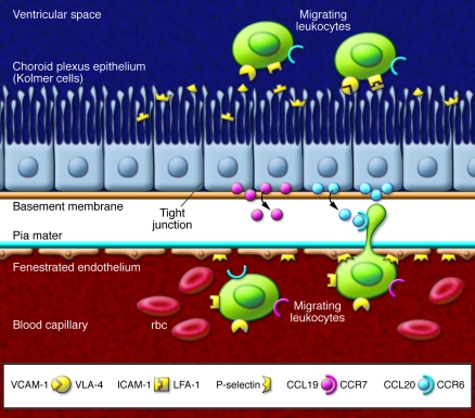
The choroid plexus is composed of highly invaginated loops of capillaries and pia
mater that reach into the ventricles of the brain. Cells from the blood and under
the influence of chemokines undergo adhesion, rolling, and diapedesis across the
fenestrated capillary endothelium and pia mater of the choroid plexus. The
basement membrane and tight junctions of the choroid plexus epithelium provide a
further barrier, the brain-CSF barrier. These modified epithelial cells (Kolmer
cells) have bulbous microvilli that secrete the CSF. Infiltrating leukocytes
migrating through these cells enter the ventricles that contain CSF and circulate
around the CNS. The chemokine CCL20 is expressed on the basolateral side of the
choroid plexus epithelial cells, attracting CCR6-expressing CD4+ T
cells. Chemokines and their receptors demonstrated to be involved in the
trafficking of immune cells into the CSF are provided in Table 2.
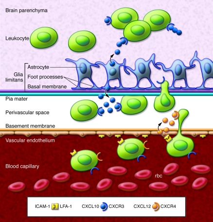
Activated leukocytes expressing adhesion molecules and integrins roll and attach
to the vascular endothelium. Successful diapedesis requires appropriate ligation
of adhesion molecules, selectins, and integrins, signaling to both the
infiltrating leukocyte and the brain endothelium. Expression of CXCL12 on the
basolateral surface of endothelial cells recruits CXCR4+ T cells.
However, retention of cells in the perivascular space occurs in the presence of
high concentrations of CXCL10. Continued migration puts cells in contact with the
glia limitans, which is composed of a highly structured wall of astrocytes.
Further positive migratory signals, including chemokines, from these and
surrounding cells may allow leukocyte migration into the parenchyma.
References
-
- Ehrlich P.Das sauerstufbudurfnis des organismus. Eine Farbenanalytische Studie . Berlin, Germany: Hirschwald; 1885.
-
- Goldmann EE. Vitalfarbung am zentralnervensystem. Abhandl Konigl preuss Akad Wiss. 1913;1:1–60.
-
- Ge S, Song L, Serwanski DR, Kuziel WA, Pachter JS. Transcellular transport of CCL2 across brain microvascular endothelial cells. J Neurochem. 2008;104(5):1219–1232. - PubMed
Publication types
MeSH terms
Substances
Grants and funding
LinkOut - more resources
Full Text Sources
Other Literature Sources
Medical
