. 2010 Aug;90(8):1199-208.
doi: 10.1038/labinvest.2010.88. Epub 2010 May 3.

Hepatic stellate cells' involvement in progenitor-mediated liver regeneration

Affiliations

Hepatic stellate cells' involvement in progenitor-mediated liver regeneration

Dana G Pintilie et al. Lab Invest. 2010 Aug.

Abstract

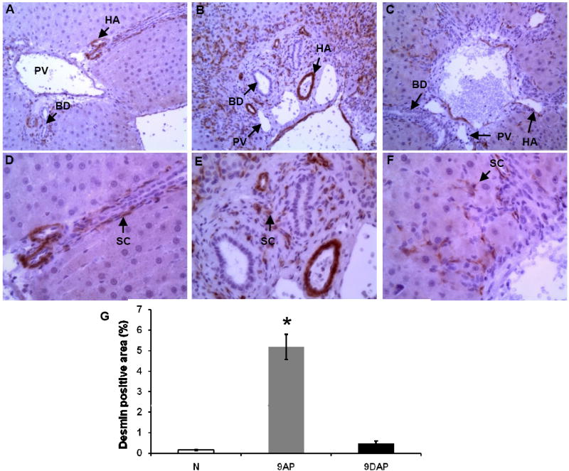
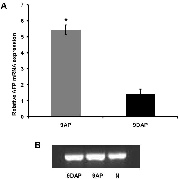
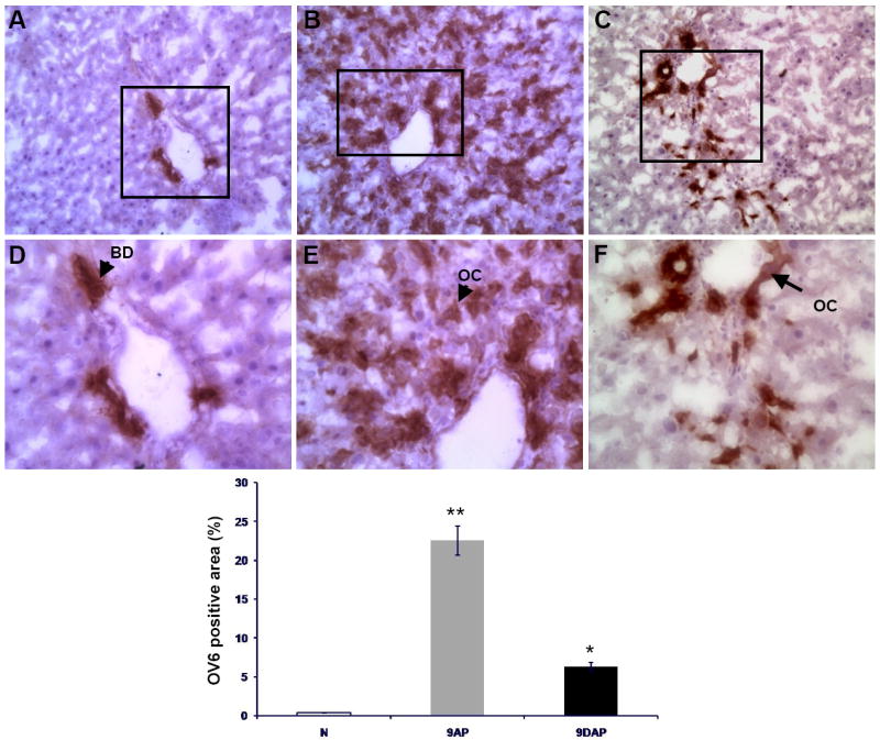
Earlier studies conducted by our laboratory have shown that suppression of transforming growth factor-beta (TGFbeta)-mediated upregulation of connective tissue growth factor (CTGF) by iloprost resulted in a greatly diminished oval cell response to 2-acetylaminofluorene/partial hepatectomy (2AAF/PH) in rats. We hypothesized that this effect is due to decreased activation of hepatic stellate cells. To test this hypothesis, we maintained rats on a diet supplemented with 2% L-cysteine as a means of inhibiting stellate cell activation during the oval cell response to 2AAF/PH. In vitro experiments show that L-cysteine did, indeed, prevent the activation of stellate cells while exerting no direct effect on oval cells. Desmin immunostaining of liver sections from 2AAF/PH animals indicated that maintenance on the L-cysteine diet resulted in an 11.1-fold decrease in the number of activated stellate cells within the periportal zones. The total number of cells proliferating in the periportal zones of livers from animals treated with L-cysteine was drastically reduced. Further analyses showed a greater than fourfold decrease in the magnitude of the oval cell response in animals maintained on the L-cysteine diet as determined by immunostaining for both OV6 and alpha-fetoprotein (AFP). Global liver expression of AFP as measured by real-time PCR was shown to be decreased 4.7-fold in the L-cysteine-treated animals. These data indicate that the activation of hepatic stellate cells is required for an appropriate oval cell response to 2AAF/PH.


Figures

Figure 1
Figure 1
Study design – diagrammatic representation of the hepatic stellate cell inhibition and oval cell induction model in ♂ Fisher 344 rats: 2% L-cysteine diet, 2-AAF pellet implantation, partial hepatectomy and selected time points for sacrifice (mo – month, w – week, d – day).
Figure 2
Figure 2
In vitro effects of L-cysteine on proliferation of selected hepatic cell populations: (A, D) WB F344 cells, (B,E) primary portal fibroblasts, (C,F) HSC T6 cells. Panels A-C received no treatment while panels D-F were cultured in media supplemented with 2% L-cysteine; all shown at x10 magnification.
Figure 3
Figure 3
BrdU index: quantitative analysis of L-cysteine effects on cultured mesenchymal and oval cells’ proliferation: WB 344F – rat oval cell line; PF – primary isolated portal fibroblasts; HSC T6 – hepatic stellate cell line; white columns – cells treated with 100 μM L-cysteine in culture medium; black columns – cells grown on culture medium w/out L-cysteine supplement.
Figure 4
Figure 4
Comparative histological exam of Hematoxylin & Eosin stained liver samples shows differences in the hepatic regeneration profile of L-cysteine fed animals: (A, D) normal animals, (B, E) 2-AAF/PH treated rats 9 days post PH, (C, F) L-cysteine/2-AAF/PH protocol 9 days after acute liver injury. BD – bile duct, PV – portal vein branch, HA – hepatic artery branch, OC – oval cell, SHL – small hepatocyte-like cell. Upper panels are shown at x20, lower panels at x40 magnification.
Figure 5
Figure 5
In vivo reduced activation of hepatic stellate cell population indicated by desmin immunostaining of liver sections from L-cysteine-fed rats: normal rat liver (A, D, N), liver at day 9 following 2AAF/PH (B, E, 9AP), same time point liver section from animals subjected to L-cysteine diet/2AAF/PH (C, F, 9DAP). BD – bile duct, PV – portal vein branch, HA – hepatic artery branch, SC – stellate cell. Upper panels are shown at x20, lower panels at x40 magnification. (G) Quantitative computer image analysis of desmin positive areas on the immunostained liver samples confirms the significant reduction of stellate cell activation on immunostained sections.
Figure 6
Figure 6
Assessment or proliferation status of hepatic cell populations by quantitative analysis of Ki67 positive nuclei on random microscopic fields: normal liver (A, N) and samples collected 9 days after the acute liver injury; (B, 9AP) rats subjected to the 2AAF/PH protocol, (C, 9DAP) animals fed with L-cysteine during the oval cell activation treatment. All shown at x20 magnification.
Figure 7
Figure 7
Concomitant reductions in proliferation of hepatic cell populations and oval cell activation, reflected by Ki67 and desmin immunofluorencent staining, respectively: normal liver (A, D); sections collected 9 days post hepatectomy from animals subjected to 2AAF/PH protocol (B, E) and rats maintained on L-cysteine/2AAF/PH (C, F). PV – portal vein branch, HA – hepatic artery branch. Upper panels are shown at x20, lower panels at x40 magnification.
Figure 8
Figure 8
Reduced oval cell presence on AFP immunostained liver sections suggested by quantitative image analysis (G): normal liver (A,D, N), 2AAF/PH treated rats day 9 following PH (B,E, 9AP) and 2AAF/PH treated rats maintained on the 2% L-cysteine diet 9 days following PH (C,F, 9DAP), OC – oval cell, TC – transitional hepatocytes. Upper panels are shown at x10, lower panels at x20 magnification.
Figure 9
Figure 9
Magnitude of oval cell response quantified by real time PCR analysis of AFP mRNA, relative to normal liver tissue expression and normalized to beta actin (A), 9AP – animals fed normal rat chow and subjected to 2AAF/PH protocol; 9DAP – animals maintained on the L-cysteine diet associated to 2AAF/PH treatment. GAPDH was used as a loading control (B).
Figure 10
Figure 10
Oval cell response reduction assessed by quantitative image analysis (D) of OV6 positive areas on tissue sections obtained from normal liver (A, N) and animals sacrificed 9 days after the acute liver injury: (B, 9AP) subjected to the 2-AAF/PHx protocol, and (C, 9DAP) fed L-cysteine during the oval cell activation treatment. OC – oval cell, BD – bile duct. Upper panels are shown at x20, lower panels at x40 magnification.

References

    1. Kuwahara R, Kofman AV, Landis CS, Swenson ES, Barendswaard E, Theise ND. The hepatic stem cell niche: identification by label-retaining cell assay. Hepatology. 2008;47(6):1994–2002. - PMC - PubMed
    1. Xiao JC, Jin XL, Ruck P, Adam A, Kaiserling E. Hepatic progenitor cells in human liver cirrhosis: immunohistochemical, electron microscopic and immunofluorencence confocal microscopic findings. World J Gastroenterol. 2004;10(8):1208–11. - PMC - PubMed
    1. Pi L, Oh SH, Shupe T, Petersen BE. Role of connective tissue growth factor in oval cell response during liver regeneration after 2-AAF/PHx in rats. Gastroenterology. 2005;128(7):2077–88. - PMC - PubMed
    1. Friedman SL. Liver fibrosis – from bench to bedside. J Hepatol. 2003;38(Suppl 1):S38–S53. - PubMed
    1. Ujike K, Shinji T, Hirasaki S, Shiraha H, Nakamura M, Tsuji T, Koide N. Kinetics of expression of connective tissue growth factor gene during liver regeneration after partial hepatectomy and D-galactosamine-induced liver injury in rats. Biochem Biophys Res Commun. 2000;277:448–454. - PubMed

Publication types

MeSH terms