. 2010 Jul;152(7):1117-27.
doi: 10.1007/s00701-010-0636-3. Epub 2010 May 4.

Mechanisms of cerebellar tonsil herniation in patients with Chiari malformations as guide to clinical management

Affiliations

Mechanisms of cerebellar tonsil herniation in patients with Chiari malformations as guide to clinical management

Thomas H Milhorat et al. Acta Neurochir (Wien). 2010 Jul.

Abstract

Background: The pathogenesis of Chiari malformations is incompletely understood. We tested the hypothesis that different etiologies have different mechanisms of cerebellar tonsil herniation (CTH), as revealed by posterior cranial fossa (PCF) morphology.

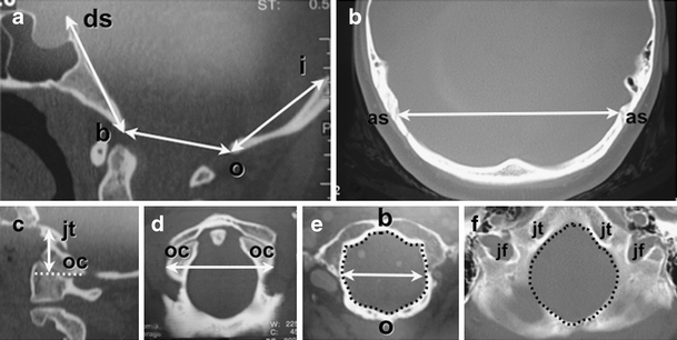
Methods: In 741 patients with Chiari malformation type I (CM-I) and 11 patients with Chiari malformation type II (CM-II), the size of the occipital enchondrium and volume of the PCF (PCFV) were measured on reconstructed 2D-CT and MR images of the skull. Measurements were compared with those in 80 age- and sex-matched healthy control individuals, and the results were correlated with clinical findings.

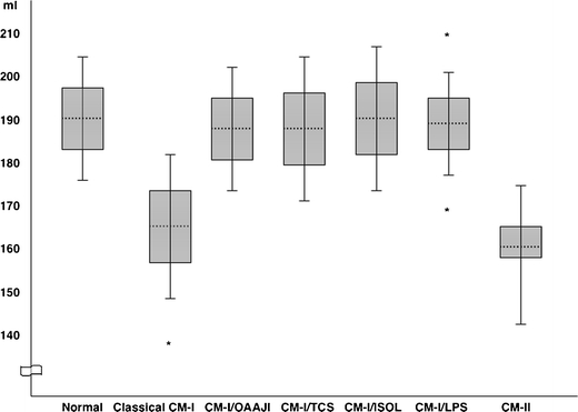
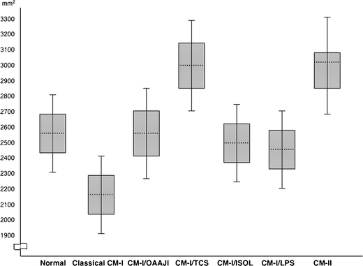
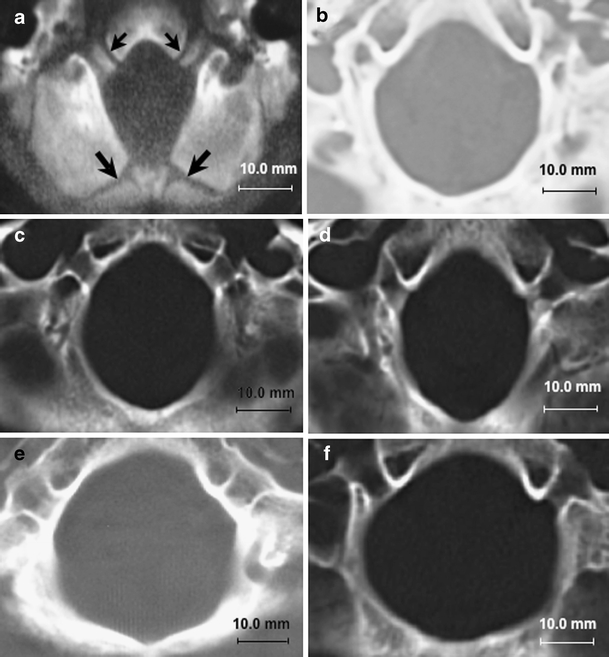
Results: Significant reductions of PCF size and volume were present in 388 patients with classical CM-I, 11 patients with CM-II, and five patients with CM-I and craniosynostosis. Occipital bone size and PCFV were normal in 225 patients with CM-I and occipitoatlantoaxial joint instability, 55 patients with CM-I and tethered cord syndrome (TCS), 30 patients with CM-I and intracranial mass lesions, and 28 patients with CM-I and lumboperitoneal shunts. Ten patients had miscellaneous etiologies. The size and area of the foramen magnum were significantly smaller in patients with classical CM-I and CM-I occurring with craniosynostosis and significantly larger in patients with CM-II and CM-I occurring with TCS.

Conclusions: Important clues concerning the pathogenesis of CTH were provided by morphometric measurements of the PCF. When these assessments were correlated with etiological factors, the following causal mechanisms were suggested: (1) cranial constriction; (2) cranial settling; (3) spinal cord tethering; (4) intracranial hypertension; and (5) intraspinal hypotension.


Conflict of interest statement

None

Figures

Fig. 1
Fig. 1
Morphometric analysis of PCF using reconstructed 2D-CT images in 25-year-old healthy control female. The size of the occipital bone was determined by measuring its enchondral parts (exocciput, basiocciput, and supraocciput) which enclose the posterior cranial fossa. a Axial length of clivus (basiocciput and basisphenoid) measured from the top of the dorsum sellae (ds) to the basion (b); axial length of supraocciput measured from the center of the internal occipital protuberance (i) to the opisthion (o); and anterior–posterior diameter of foramen magnum measured as the distance between the inner surfaces of the basion and opisthion on midsaggital images. b Width of supraocciput measured as the distance between the inner surfaces of the asterions (as). c The axial height of occipital condyle (exocciput) was measured as the perpendicular distance between the top of the jugular tubercle (jt) and the bottom of the occipital condyle (oc) along a line parallel with the orbitomeatal line. d The width of the occipital condyles (oc) was measured as the distance between their outer surfaces. e The maximum transverse diameter of the inferior outlet of the foramen magnum was measured at the level of the basion (b) and opisthion (o). The area of the inferior outlet of the foramen magnum is enclosed by dotted line. f The area of the superior outlet of the foramen magnum was measured at level of the jugular tubercles (jt) and jugular foramina (jf) and is enclosed by dotted line
Fig. 2
Fig. 2
Ranges of PCFV in patients with Chiari malformations stratified by etiology. In patients with CM-I, means are shown by dotted lines; 1 SD ranges are shown by boxes, and 2 SD ranges are shown by whiskers. In patients with CM-II, the median is shown by dotted line; the interquartile range is shown by box, and the maximum/minimum ranges are shown by whiskers owing to non-Gaussian distribution. Asterisks indicate three patients with PCFV measurements greater than 2 SD. Chiari subgroups with less than ten patients are not represented. Abbreviations: CM-I Chiari malformation type I, OAAJI occipitoatlantoaxial joint instability, TCS tethered cord syndrome, ISOL intracranial space-occupying lesions, LPS lumboperitoneal shunt, CM-II Chiari malformation type II
Fig. 3
Fig. 3
Ranges of summated inferior and superior foramen magnum outlet areas stratified by etiology. In patients with CM-I, means are shown by dotted lines, 1 SD ranges are shown by boxes, and 2SD ranges are shown by whiskers. In patients with CM-II, the median is shown by dotted line; the interquartile range is shown by box, and the maximum/minimum ranges are shown by whiskers owing to non-Gaussian distribution. Chiari subgroups with less than ten patients are not represented. Abbreviations: CM-I Chiari malformation type I, OAAJI occipitoatlantoaxial joint instability, TCS tethered cord syndrome, ISOL intracranial space-occupying lesions, LPS lumboperitoneal shunt, CM-II Chiari malformation type II
Fig. 4
Fig. 4
Morphometric assessments of foramen magnum at level of superior outlet using axial CT images. a Normal foramen magnum in 10-month-old male showing patent basi-exoccipital (small arrows) and exosupraoccipital synchondroses (large arrows). b Normal foramen magnum in 30-year-old female with anterior–posterior diameter of 34.2 mm, transverse diameter of 31.0 mm, and area of 1,872.4 mm2. c Abnormally small foramen magnum in 28-year-old female with classical CM-I. There are significant reductions of the transverse diameter (25.2 mm) and area (1,187.5 mm2). The anterior–posterior diameter is normal giving the foramen magnum an exaggerated oval shape. d Abnormally small foramen magnum in 25-year-old female with CM-I and craniosynostosis (Crouzon’s disease). There are significant reductions of the transverse diameter of the foramen magnum (24.8 mm) and area (1,185.4 mm2). The anterior–posterior diameter is normal giving the foramen magnum an exaggerated oval shape. e Abnormally large foramen magnum in 27-year-old female with CM-I and tethered cord syndrome. There are significant increases of the anterior–posterior diameter (40.5 mm), transverse diameter (35.1 mm), and area (2,084.7 mm2) giving the foramen magnum a rounded shape. f Abnormally large foramen magnum in 17-year-old male with CM-II. There are significant increases of the anterior–posterior (37.5 mm), transverse diameter (37.1 mm), and area (2,087.7 mm2) giving the foramen magnum a rounded shape. Abbreviations: CM-I Chiari malformation type I, CM-II Chiari malformation type II

References

    1. Abel TJ, Chowdhary A, Gabikian P, Ellenbogen RG, Avellino AM. Acquired Chiari malformation type I associated with a fatty terminal filum. Case report. J Neurosurg. 2006;105:329–332. - PubMed
    1. Atkinson JL, Weinshenker BG, Miller GM, Piegras DG, Mokri B. Acquired Chiari I malformation secondary to spontaneous spinal cerebrospinal fluid leakage and chronic intracranial hypotension syndrome in seven cases. J Neurosurg. 1998;88:237–242. doi: 10.3171/jns.1998.88.2.0237. - DOI - PubMed
    1. Barry A, Patten BM, Stewart BH. Possible factors in the development of the Arnold–Chiari malformation. J Neurosurg. 1957;14:285–301. doi: 10.3171/jns.1957.14.3.0285. - DOI - PubMed
    1. Chiari H. Über Veränderungen des Kleinhirns in folge von Hydrocephalie des Grosshirns. Dtsch Med Wochenschr. 1891;17:1172–11725. doi: 10.1055/s-0029-1206803. - DOI
    1. Chumas PD, Armstrong DC, Drake JM, Kulkarni AV, Hoffman HJ, Rutka HRP, JT HEB. Tonsillar herniation: the rule rather than the exception after lumboperitoneal shunting in the pediatric population. J Neurosurg. 1993;78:568–573. doi: 10.3171/jns.1993.78.4.0568. - DOI - PubMed

Publication types