Review
. 2010 Jun;45(3):171-84.
doi: 10.3109/10409238.2010.480968.

A new model for SOS-induced mutagenesis: how RecA protein activates DNA polymerase V

Affiliations
Review

A new model for SOS-induced mutagenesis: how RecA protein activates DNA polymerase V

Meghna Patel et al. Crit Rev Biochem Mol Biol. 2010 Jun.

Abstract

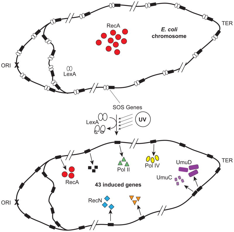
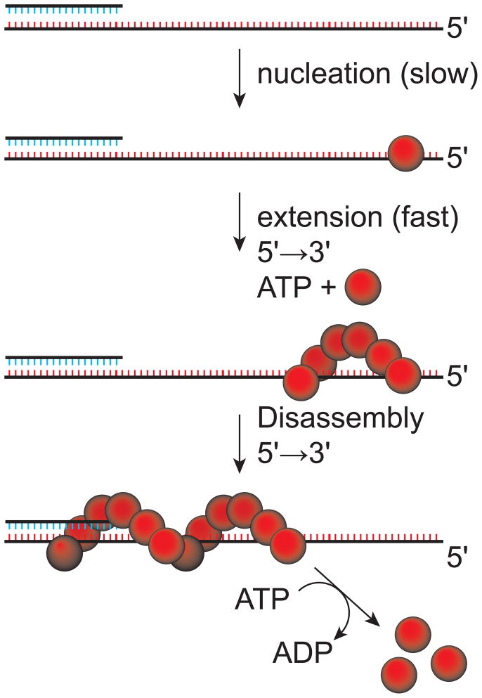
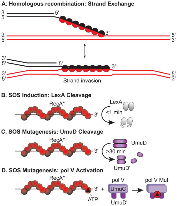
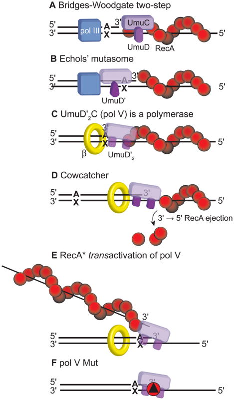
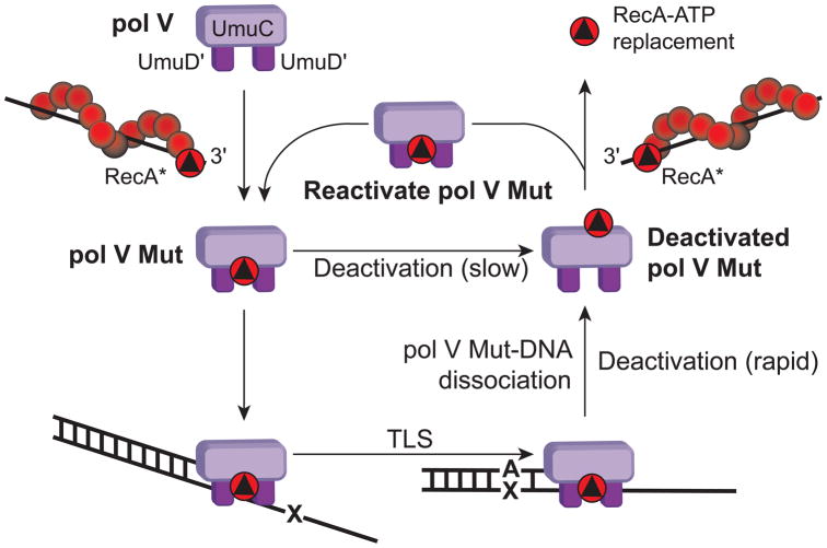
In Escherichia coli, cell survival and genomic stability after UV radiation depends on repair mechanisms induced as part of the SOS response to DNA damage. The early phase of the SOS response is mostly dominated by accurate DNA repair, while the later phase is characterized with elevated mutation levels caused by error-prone DNA replication. SOS mutagenesis is largely the result of the action of DNA polymerase V (pol V), which has the ability to insert nucleotides opposite various DNA lesions in a process termed translesion DNA synthesis (TLS). Pol V is a low-fidelity polymerase that is composed of UmuD'(2)C and is encoded by the umuDC operon. Pol V is strictly regulated in the cell so as to avoid genomic mutation overload. RecA nucleoprotein filaments (RecA*), formed by RecA binding to single-stranded DNA with ATP, are essential for pol V-catalyzed TLS both in vivo and in vitro. This review focuses on recent studies addressing the protein composition of active DNA polymerase V, and the role of RecA protein in activating this enzyme. Based on unforeseen properties of RecA*, we describe a new model for pol V-catalyzed SOS-induced mutagenesis.


Figures

Figure 1
Figure 1
The SOS response in Escherichia coli. The system includes at least 43 known genes. Only a subset of these are shown. Under normal growth conditions, the SOS genes are repressed by the dimeric transcriptional repressor, LexA. Upon exposure to DNA damage, such as ultraviolet radiation, the LexA repressor is autocatalytically cleaved in a reaction involving the RecA protein, and the SOS genes are induced.
Figure 2
Figure 2
Assembly and disassembly of RecA protein filaments. RecA filament nucleation is the slow step, normally occurs on single-stranded DNA, requires ATP, and involves an oligomer with 4–5 RecA subunits in it. Extension of the filament occurs rapidly in the 5′ → 3′ direction by addition of RecA subunits. Extension will continue into adjacent regions of double stranded DNA. Disassembly also occurs in the 5′ → 3′ direction, and from the opposite end of the filament. Disassembly is coupled to ATP hydrolysis. RecA filaments formed on any DNA in the presence of ATP are active in activating DNA polymerase V, and have been designated RecA*.
Figure 3
Figure 3
The four cellular functions of the RecA protein in Escherichia coli. (A) RecA protein is involved in many aspects of recombinational DNA repair, and promotes a variety of DNA strand exchange reactions in the context of this function. One key type of reaction is shown – DNA strand invasion. In this process, RecA forms a filament (RecA*) on the 3′ end of a single-stranded DNA, aligns that DNA with its complement in a duplex DNA, and pairs the bound DNA with the complementary strand of the duplex (displacing the other duplex strand). (B) The autocatalytic cleavage of LexA repressor occurs rapidly in the presence of RecA* filaments. It is the number and extent of filament formation that governs the efficiency of LexA auto cleavage. As a result of excessive DNA damage, the number of single strand DNA gaps are increased. LexA cleavage and the accompanying SOS system induction generally occur only in response to such damage. (C) RecA* is required for the autocatalytic cleavage of the UmuD protein to form active UmuD′. This reaction is much slower than LexA cleavage and occurs >30mins after DNA damage. (D) A RecA monomer and a molecule of ATP are transferred to pol V from the 3′-proximal end of a RecA* filament to form the active pol V Mut.
Figure 4
Figure 4
Translesion synthesis (TLS) models. (A) The Bridges–Woodgate two-step model. In this model, translesion synthesis (TLS) is catalysed by DNA polymerase (pol) III. DNA pol III (which requires RecA in a first step) inserts a nucleotide opposite a template lesion (for example, A opposite X) and, in a second step, also copies past the lesion, which requires the UV mutagenesis gene products UmuDC. (B) The Echols mutasome model. A multiprotein complex that includes UmuC, UmuD′ and DNA pol III holoenzyme is recruited to a DNA lesion by a RecA nucleoprotein filament. The mutasome complex enables replication to take place across the lesion, which results in mutations. (C) UmuD′2C (pol V) is a DNA polymerase. An in vitro system composed of UmuD′2C, RecA and the β-sliding clamp carries out TLS in the absence of DNA pol III. The UmuD′2C complex (designated DNA pol V) was shown to have intrinsic DNA polymerase activity in its UmuC subunit. (D) The cowcatcher model. The presence of a RecA nucleoprotein filament proximal to a lesion blocks TLS that is mediated by DNA pol V. Analogous to a cowcatcher on the front of a locomotive, DNA pol V in the presence of single-stranded binding protein (SSB, not shown) removes RecA in a 3′→5′ direction ahead of the advancing DNA pol V to allow TLS to occur. (E) RecA* transactivation of DNA pol V model. TLS requires DNA pol V to be activated by interacting with the 3′-proximal tip of RecA bound to a separate single-stranded (ss) DNA molecule in trans. A proficient transactivating RecA nucleoprotein filament is formed on gapped DNA. (F) Pol V Mut model. RecA* is required to directly activate pol V by transferring a molecule of RecA (‘red circle’ and ATP ‘black triangle’ from the 3′-proximal tip of RecA* to form an activated pol V mutasome, UmuD′2C-RecA-ATP, which catalyses TLS in the absence of RecA*.
Figure 5
Figure 5
“Fly in the cis RecA* filament ointment”. (A) Pol V (UmuD′2C) synthesis and TLS measured on a 64 nt ssDNA template overhang containing an abasic lesion, X, located 50 nt from the 5′-template end, in the presence of ATPγS, SSB, and different RecA/DNA nt ratios, as indicated. (B) Primer utilization (●) and lesion bypass efficiencies (○) calculated from the data in Figure 5A. Although a much more robust lesion bypass reaction occurs when conditions are optimal for filament assembly with ~1 RecA molecule per 3 nucleotides, Pol-V-catalyzed TLS is clearly observed at a stoichiometry of ~1 RecA molecule per 50 nucleotides, conditions under which RecA* formation is absent.
Figure 6
Figure 6
Kinetics of primer/template annealing. When forming primer/template DNA molecules by mixing single-stranded DNA primers and templates and then allowing them to anneal in a bimolecular reaction, the presence of ssDNA on which a RecA filament can assemble cannot be avoided. There will always be un-annealed template and primer DNA and therefore, an indeterminate amount of single stranded DNA in all experiments utilizing annealed primer/template substrates. A simple way to eliminate excess ssDNA is to form a stable hairpin structure so that almost all of the DNA anneals in the form of a hairpin, in a unimolecular reaction, with a very small proportion of non-hairpin ssDNA remaining. By having a short template overhang (3 nt), RecA filament assembly on the hairpin primer/template DNA is eliminated, because the footprint of a single RecA monomer is 3 nt.
Figure 7
Figure 7
Pol V transactivation by RecA*. Pol V-catalysed primer utilization when copying a hairpin containing a 3-nucleotide-long template overhang was measured in the presence of ATPγS and RecA (2 μM) at varying trans ssDNA (36-mer) concentrations (in nM). The specific activity of pol V is measured in pmol DNA per μg pol V per min. The arrowhead indicates the position of the full-length product. ‘%PU’ represents the fraction of primer utilized, i.e., extended by at least 1 nt.
Figure 8
Figure 8
Determination of the molecular mass of pol V Mut by MALS. In the upper panel, after RecA*-mediated transactivation of UmuD′2C and removal of RecA*, the mixture of pol V Mut and non-activated pol V was resolved by size-exclusion chromatography (upper trace), and the molecular mass corresponding to each peak was measured by multiangle light scattering (MALS). Non-activated pol V run separately on the silica gel elutes at 18.4 min (lower trace). In the lower panel, silver-stained SDS–polyacrylamide gel shows the protein composition from the two peaks contained in the upper panel (upper trace).
Figure 9
Figure 9
TLS performed by pol V Mut in the absence of RecA*. DNA template lesions, a cis-syn TT dimer and an abasic moiety, are copied by pol V Mut-RecA WT or pol V Mut-RecA E38K/ΔC17 in the absence of preformed trans RecA* filaments. Without preactivation, pol V is virtually inactive. “N” refers to a normal undamaged DNA template strand. “X” denotes a lesion. The primer strand is 12 nucleotides long and the template overhang is 36 nucleotides long.
Figure 10
Figure 10
Pol V Mut non-cycling. Each active pol V Mut complex can promote only one round of DNA synthesis, after which it cannot reinitiate synthesis on a different p/t DNA, and is thus deactivated (●). The deactivated complex can be fully resurrected by addition of trans RecAE38K/ΔC17*, which enables pol V Mut to reinitiate DNA synthesis on a separate p/t DNA substrate and facilitate cycling of the enzyme (▼). Addition of free RecAE38K/ΔC17 does not reinitiate DNA synthesis (■). Trans RecA* is at 1 μM when present. The reactions are performed under conditions of excess substrate DNA to active enzyme.
Figure 11
Figure 11
Model for pol V Mut function. Pol V is UmuD′2C, and is minimally active on its own. Transfer of an ATP-bound RecA subunit from RecA* creates the active pol V Mut. Pol V Mut can migrate to a template-primer site where its activity is required. There, it will extend the primer and insert nucleotides opposite any lesion encountered (called translesion synthesis or TLS). Upon dissociation, pol V Mut is inactivated. There is also a slow inactivation of pol V Mut in solution, without carrying out TLS. Deactivated pol V Mut can be reactivated by interaction with another RecA* filament, with transfer of a new RecA-ATP subunit and displacement of the old RecA-ATP subunit.

Similar articles

Cited by

References

    1. Arenson TA, Tsodikov OV, Cox MM. Quantitative analysis of the kinetics of end-dependent disassembly of RecA filaments from ssDNA. J Mol Biol. 1999;288:391–401. - PubMed
    1. Bienko M, Green CM, Crosetto N, Rudolf F, Zapart G, Coull B, Kannouche P, Wider G, Peter M, Lehmann AR, Hofmann K, Dikic I. Ubiquitin-binding domains in Y-family polymerases regulate translesion synthesis. Science. 2005;310:1821–4. - PubMed
    1. Bishop DK, Park D, Xu L, Kleckner N. DMC1: a meiosis-specific yeast homolog of E. coli recA required for recombination, synaptonemal complex formation, and cell cycle progression. Cell. 1992;69:439–56. - PubMed
    1. Bork JM, Cox MM, Inman RB. RecA protein filaments disassemble in the 5′ to 3′ direction on single-stranded DNA. Journal of Biological Chemistry. 2001;276:45740–43. - PubMed
    1. Boudsocq F, Campbell M, Devoret R, Bailone A. Quantitation of the inhibition of Hfr × F- recombination by the mutagenesis complex UmuD′C. J Mol Biol. 1997;270:201–11. - PubMed

Publication types

MeSH terms

LinkOut - more resources