. 2010 Aug 1;49(3):383-91.
doi: 10.1016/j.freeradbiomed.2010.04.026. Epub 2010 May 2.

Accumulation of oxidized proteins in Herpesvirus infected cells

Affiliations

Accumulation of oxidized proteins in Herpesvirus infected cells

Shomita S Mathew et al. Free Radic Biol Med. .

Abstract

Oxidative stress gives rise to an environment that can be highly damaging to proteins, lipids, and DNA. Previous studies indicate that Herpesvirus infections cause oxidative stress in cells and in tissues. The biological consequences of virus-induced oxidative stress have not been characterized. Studies from many groups indicate that proteins which have been damaged through oxidative imbalances are either degraded by the 20S proteasome in a ubiquitin-independent fashion or form aggregates that are resistant to proteolysis. We have previously shown that herpes simplex virus type 1 (HSV-1) replication was significantly enhanced in the presence of the cellular antioxidant chaperone Hsp27, indicating a possible role for this protein in managing virus-induced oxidative stress. Here we show that oxidized proteins accumulate during infections with two distantly related herpesviruses, HSV-1 and Rhesus Rhadinovirus (RRV), a close relative of the Kaposi's sarcoma-associated herpesvirus (KSHV). The presence of oxidized proteins was not entirely unexpected as oxidative stress during herpesvirus infection has been previously documented. Unexpectedly, some oxidized proteins are removed in a proteasome-dependent fashion throughout infection and others resist degradation. Oxidized proteins that resist proteolysis become sequestered in foci within the nucleus and are not associated with virus-induced chaperone enriched domains (VICE), active centers of protein quality control, but rather coincide with Hsp27-enriched foci that were previously described by our laboratory. Experiments also indicate that the accumulation of oxidized proteins is more pronounced in cells depleted for Hsp27. We propose that Hsp27 may facilitate oxidized protein turnover at VICE domains in the nucleus during infection. Hsp27 may also buffer toxic effects of highly-carbonylated, defective proteins that resist proteolysis by promoting their aggregation in the nucleus. These roles of Hsp27 during virus infection are most likely not mutually exclusive.


Figures

Figure 1
Figure 1
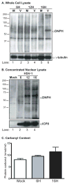
DNPH levels in HSV-1 infected Vero cells. A. Western blot analysis of DNPH derived proteins in whole cell lysates of Vero cells mock-infected (M) or infected (V) with HSV-1 at 6, 12 and 16 hours post infection (hpi). B. Western blot analysis of DNPH derived proteins in concentrated nuclear lysates of Vero cells mock infected or infected with HSV-1 at 6, 12 and 16 hpi. γ-tubulin is included as the loading control for these assays and viral ICP8 is the infection control. C. Graphical representation of protein carbonyl content in mock-infected and HSV-1 infected (6 and 16 hours) Vero cells as measured by carbonyl content assay. The migration patterns of protein molecular weight standards are indicated.
Figure 2
Figure 2
DNPH levels and protein carbonyl content in Rhesus Rhadinovirus (RRV-GFP) infected Rhesus macaque fibroblasts. A. Western blot analysis of the levels of DNPH derived proteins in fibroblasts infected with RRV-GFP at an MOI of 1 for 1 and 5 days. L26 is included as the loading control for these assays and GFP expression is used to gauge productive infection. The progression of infection was monitored by GFP expression. A light and dark exposure of the Western blot are provided. The migration patterns of protein molecular weight standards are indicated. B. Graphical representation of protein carbonyl content in mock-infected and RRV-GFP infected (1 and 5 days) fibroblasts as measured by the carbonyl content assay.
Figure 3
Figure 3
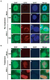
Distribution of DNPH derived proteins in HSV-1- infected Vero cells. A. Conventional immunofluorescence analysis of Vero cells that were either mock-infected (a–h) or infected with HSV-1 (i–p). B. Denaturing immunofluorescence analysis of Vero cells that were mock-infected (a–d) or infected with HSV-1 (e–l). Shown are staining profiles for DNP (green), viral DNA binding protein ICP8 (red) and DAPI (blue). The three signals are shown merged in the last column.
Figure 4
Figure 4
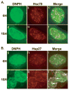
Distribution of DNPH and the cellular chaperone proteins Hsc70 and Hsp27 in HSV-1 infected Vero cells. A. Denaturing immunofluorescence analysis on Vero cells infected with HSV-1 for 6h (a–c) or 16h (d–f). Staining profiles are shown for DNPH (green) and the cellular chaperone protein Hsc70 (marker for VICE domains). The two signals are shown merged in the last column. B. Denaturing immunofluorescence analysis of Vero cells infected for 6h (g–i) or 16h (j–l) and stained for DNPH (green) and Hsp27 (red). Merged images for the two signals are shown in the last column.
Figure 5
Figure 5
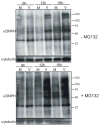
A. Western blot analysis of DNPH derived proteins in untreated Vero cells mock infected (M) or infected (V) with HSV-1 at 6, 12 and 16 hpi. B. Western blot analysis of DNPH derived proteins in MG132-treated Vero cells mock infected (M) or infected (V) with HSV-1 at 6, 12 and 16 hpi. MG132 was added (+) at 6 hpi. γ-tubulin is included as the loading control for these assays. The migration patterns of protein molecular weight standards are indicated.
Figure 6
Figure 6
Depletion of Hsp27 with siRNA technology and carbonyl content of HSV-1 infected HeLa cells. A. Western blot analysis of Hsp27, viral marker for infection (ICP8) and a loading control (γ-tubulin) in cells treated with a non-targeting siRNA (lanes 1–3) and Hsp27-specific siRNA (lanes 4–6). B. Graphical representation of protein carbonyl content measured using a carbonyl content assay in HeLa cells treated with non targeting (NT si) or hsp27-specific (H27 si) siRNA and either mock-infected or HSV-1 infected for 6 and 16h.

Similar articles

Cited by

References

    1. Burch AD, Weller SK. Nuclear sequestration of cellular chaperone and proteasomal machinery during herpes simplex virus type 1 infection. J Virol. 2004;78:7175–7185. - PMC - PubMed
    1. Burch AD, Weller SK. Herpes simplex virus type 1 DNA polymerase requires the mammalian chaperone hsp90 for proper localization to the nucleus. J Virol. 2005;79:10740–10749. - PMC - PubMed
    1. Livingston CM, Ifrim MF, Cowan AE, Weller SK. Virus-Induced Chaperone-Enriched (VICE) domains function as nuclear protein quality control centers during HSV-1 infection. PLoS Pathog. 2009;5:e1000619. - PMC - PubMed
    1. Li L, Johnson LA, Dai-Ju JQ, Sandri-Goldin RM. Hsc70 focus formation at the periphery of HSV-1 transcription sites requires ICP27. PLoS ONE. 2008;3:e1491. - PMC - PubMed
    1. Mathew SS, Della Selva MP, Burch AD. Modification and reorganization of the cytoprotective cellular chaperone Hsp27 during herpes simplex virus type 1 infection. J Virol. 2009;83:9304–9312. - PMC - PubMed

Publication types