. 2010 Jul-Aug;3(7-8):471-85.
doi: 10.1242/dmm.004598. Epub 2010 May 4.

Fragile X mental retardation protein has a unique, evolutionarily conserved neuronal function not shared with FXR1P or FXR2P

Affiliations

Fragile X mental retardation protein has a unique, evolutionarily conserved neuronal function not shared with FXR1P or FXR2P

R Lane Coffee Jr et al. Dis Model Mech. 2010 Jul-Aug.

Abstract

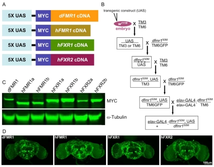
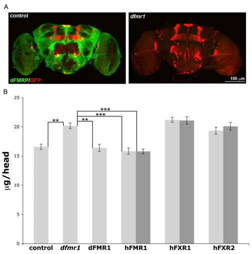
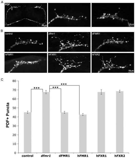
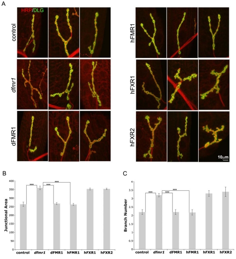
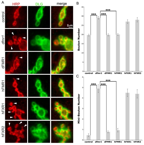
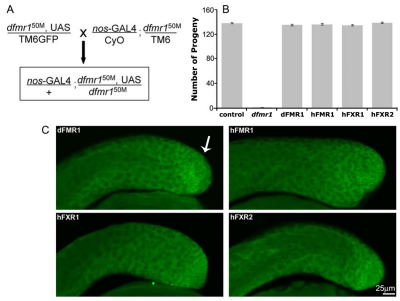
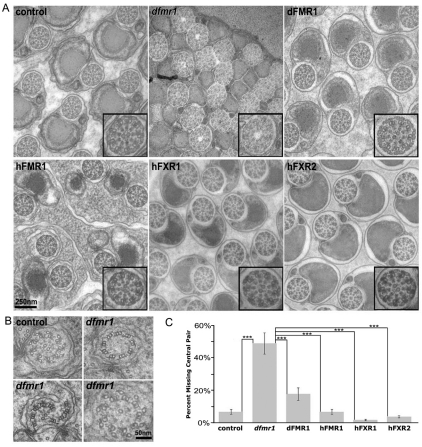
Fragile X syndrome (FXS), resulting solely from the loss of function of the human fragile X mental retardation 1 (hFMR1) gene, is the most common heritable cause of mental retardation and autism disorders, with syndromic defects also in non-neuronal tissues. In addition, the human genome encodes two closely related hFMR1 paralogs: hFXR1 and hFXR2. The Drosophila genome, by contrast, encodes a single dFMR1 gene with close sequence homology to all three human genes. Drosophila that lack the dFMR1 gene (dfmr1 null mutants) recapitulate FXS-associated molecular, cellular and behavioral phenotypes, suggesting that FMR1 function has been conserved, albeit with specific functions possibly sub-served by the expanded human gene family. To test evolutionary conservation, we used tissue-targeted transgenic expression of all three human genes in the Drosophila disease model to investigate function at (1) molecular, (2) neuronal and (3) non-neuronal levels. In neurons, dfmr1 null mutants exhibit elevated protein levels that alter the central brain and neuromuscular junction (NMJ) synaptic architecture, including an increase in synapse area, branching and bouton numbers. Importantly, hFMR1 can, comparably to dFMR1, fully rescue both the molecular and cellular defects in neurons, whereas hFXR1 and hFXR2 provide absolutely no rescue. For non-neuronal requirements, we assayed male fecundity and testes function. dfmr1 null mutants are effectively sterile owing to disruption of the 9+2 microtubule organization in the sperm tail. Importantly, all three human genes fully and equally rescue mutant fecundity and spermatogenesis defects. These results indicate that FMR1 gene function is evolutionarily conserved in neural mechanisms and cannot be compensated by either FXR1 or FXR2, but that all three proteins can substitute for each other in non-neuronal requirements. We conclude that FMR1 has a neural-specific function that is distinct from its paralogs, and that the unique FMR1 function is responsible for regulating neuronal protein expression and synaptic connectivity.


Figures

Fig. 1.
Fig. 1.
Generation of transgenic constructs with targeted neuronal expression. (A) The four UAS transgenic constructs generated and tested in this study. The positive control is wild-type dFMR1, and the three human genes are hFMR1, hFXR1 and hFXR2. All cDNA transgenic constructs are tagged with a MYC epitope in the pUAST (5X UAS) expression vector to follow protein expression. In all assays, two independent transgenic lines for each human transgenic construct were analyzed. (B) The embryonic transformation and genetic crossing scheme that was used to introduce each stably integrated UAS transgene into the dfmr1 null mutant background and then drive expression with the pan-neuronal GAL4 driver elav-GAL4. (C) Western blot analyses of transgenic protein expression for the dFMR1 line (control) and two independent lines of hFMR1, hFXR1 and hFXR2 (denoted as a/b). Expression from brain extracts (1–2-day-old adults) was tested with an anti-MYC antibody against the epitope tag common to all four transgenes (see A). Lines were selected for comparable transgene expression. The loading control is α-tubulin. (D) Brain immunohistochemistry for transgene expression of the dFMR1 line (control) and the three human lines (hFMR1, hFXR1 and hFXR2). Drosophila adult brains (1–2 days old) were probed with anti-MYC to detect the transgene epitope tag. Comparable transgene expression occurs in all conditions. Bar, 100 μm.
Fig. 2.
Fig. 2.
Only hFMR1 rescues elevated protein levels in the dfmr1 null brain. (A) Comparison of dFMRP expression in the wild-type control (w1118) and the dfmr1 null (dfmr150M) adult Drosophila brain, which were used as positive and negative controls in all assays. Acutely dissected brains (2 days old) were immunolabeled with anti-dFMRP (green) and anti-GFP (red) to reveal a transgene marker in the mushroom body learning/memory center. Note that the null mutant brain is of normal size with normal gross architecture. Bar, 100 μm. (B) Total brain protein was extracted from young adult (0–7 hours old) animals and quantified with a MicroBCA assay. The six genotypes that were compared are: w1118 control, dfmr1 null (dfmr150M), elav-GAL4 driving UASdFMR1 (positive control), and two independent lines each (light and dark gray bars) of UAS-hFMR1, UAS-hFXR1 and UAS-hFXR2 expression in the dfmr1 null background. Each bar shows the average protein levels in μg per head. Sample size: 10–20 pooled heads per sample, n=8. Significance: **P<0.01; ***P<0.001.
Fig. 3.
Fig. 3.
Only hFMR1 rescues clock neuron synapse arbors in dfmr1 null mutants. (A) Representative images of small ventrolateral (sLNv) clock neurons in the adult brain labeled with anti-PDF. The low-magnification image on the left shows the bilaterally symmetrical sLNv projections, terminating in synaptic arbor projections (arrow) in the dorsal protocerebrum. The higher magnification images show the left side (middle panel) and right side (right panel) synaptic arbors. Note the PDF-positive punctae marking the synaptic boutons. Bars, 20 μm (A, left panel); 10 μm (A, middle and right panels). (B) Representative images of the sLNv synaptic arbors from the six genotypes assayed: w1118 (control), dfmr150M null (dfmr1), and the null background with elav-GAL4-driven UAS-dFMR1, UAS-hFMR1, UAS-hFXR1 and UAS-hFXR2. Note the synaptic overgrowth characteristic of the null mutant, and the rescue of this overgrowth defect by dFMR1 and hFMR1 only. Bar, 10 μm. (C) Quantification of the number of PDF-positive punctae per synaptic arbor in the six genotypes shown. Sample size: n≥10 animals for each genotype. Significance: ***P<0.001 for all comparisons.
Fig. 4.
Fig. 4.
Only hFMR1 rescues synapse architecture in dfmr1 null mutants. The wandering third instar NMJ synapse was co-labeled with presynaptic and postsynaptic markers and compared between the six genotypes: wild-type control, dfmr1 null mutants, and elav-GAL4-driven expression in the dfmr1 null background of UAS-dFMR1 (positive control) and two independent lines each of UAS-hFMR1, UAS-hFXR1 and UAShFXR2. (A) Representative images of the muscle 4 NMJ labeled for presynaptic HRP (red) and postsynaptic DLG (green). Three example synaptic arbors are shown for each of the six genotypes. Bar, 10 μm. Quantification of junction area measured based on DLG domain expression (B) and the number of synaptic branches measured based on HRP labeling (C). The two independent lines for each human transgene were not significantly different in any case, and were therefore pooled for these comparisons. Sample size: n≥10 animals for each genotype. Significance: ***P<0.001 for all comparisons.
Fig. 5.
Fig. 5.
Only hFMR1 rescues synapse bouton differentiation in dfmr1 null mutants. (A) Representative highmagnification images of synaptic boutons. Mature type 1b boutons were defined as boutons >2 μm in minimal diameter. Satellite mini-boutons, representing an early stage in bouton differentiation, are <2 μm in diameter and are directly attached to a mature type 1b bouton (arrows). Developmentally arrested mini-boutons accumulate in the dfmr1 null mutant. Bar, 2 μm. Quantification of the number of mature boutons (B) and mini-boutons (C) per synaptic arbor in the six genotypes. The two independent lines for each human transgene were not significantly different, and were therefore pooled for these comparisons. Sample size: n≥10 animals for each genotype. Significance: ***P<0.001 for all comparisons.
Fig. 6.
Fig. 6.
All three human genes rescue dfmr1 mutant male fecundity. (A) The crossing scheme used to assay transgene function in the testes. The germline nos-GAL4 line was used to drive UAS-dFMR1 (control) and the three human transgenes (UAS-hFMR1, UAS-hFXR1 and UAS-hFXR2) in the dfmr1 null mutant background. (B) Quantification of the number of progeny per male for all six genotypes. The dfmr1 null mutant is effectively sterile owing to non-motile sperm. The two independent lines for each human transgene were not significantly different in any case, and were therefore pooled for these comparisons. (C) Representative images of adult male testes (<24 hours) with the nos-GAL4 line driving expression of the four MYC-tagged transgenes. Anti-MYC labeling was used to detect UAS-dFMR1, -hFMR1, -hFXR1 and -hFXR2. Expression was highest in the germline stem cells (arrow) and early spermatid progeny, as expected for the nos-GAL4 driver. Bar, 25 μm.
Fig. 7.
Fig. 7.
All three human genes rescue dfmr1 mutant spermatogenesis defects. (A) Representative images of the testes spermatid ultrastructure for all six genotypes. Insets show high-magnification views of a single axoneme. Wild-type sperm tails display the characteristic 9+2 microtubule arrangement of nine outer doublets and the central pair (inset). The dfmr1 null mutants exhibit disordered microtubules with the central pair missing (inset). All four transgenic lines display an ultrastructure that is indistinguishable from the wild type. Bar, 250 nm. (B) Higher magnification views of the sperm tail axoneme. Control axonemes show a perfectly arranged 9+2 microtubule organization. For dfmr1 mutants, several examples are shown displaying the range of microtubule disruption phenotypes, including the missing central pair, malformed outer ring, and milder loss of both central and outer ring microtubule integrity. Bar, 50 nm. (C) Quantification of the percentage of spermatids displaying a missing central pair of microtubules from the axoneme for all six genotypes. The two independent lines for each human transgene were not significantly different and were therefore pooled. Significance: ***P<0.001 for all comparisons.

Similar articles

Cited by

References

    1. Agulhon C, Blanchet P, Kobetz A, Marchant D, Faucon N, Sarda P, Moraine C, Sittler A, Biancalana V, Malafosse A, et al. (1999). Expression of FMR1, FXR1, and FXR2 genes in human prenatal tissues. J Neuropathol Exp Neurol. 58, 867–880 - PubMed
    1. Ashley J, Packard M, Ataman B, Budnik V. (2005). Fasciclin II signals new synapse formation through amyloid precursor protein and the scaffolding protein dX11/Mint. J Neurosci. 25, 5943–5955 - PMC - PubMed
    1. Bakker CE, de Diego Otero Y, Bontekoe C, Raghoe P, Luteijn T, Hoogeveen AT, Oostra BA, Willemsen R. (2000). Immunocytochemical and biochemical characterization of FMRP, FXR1P, and FXR2P in the mouse. Exp Cell Res. 258, 162–170 - PubMed
    1. Banerjee P, Nayar S, Hebbar S, Fox CF, Jacobs MC, Park JH, Fernandes JJ, Dockendorff TC. (2007). Substitution of critical isoleucines in the KH domains of Drosophila fragile X protein results in partial loss-of-function phenotypes. Genetics 175, 1241–1250 - PMC - PubMed
    1. Bassell GJ, Warren ST. (2008). Fragile X syndrome: loss of local mRNA regulation alters synaptic development and function. Neuron 60, 201–214 - PMC - PubMed

Publication types

MeSH terms