. 2010 Jun 1;123(Pt 11):1884-93.
doi: 10.1242/jcs.066084. Epub 2010 May 4.

Membrane wounding triggers ATP release and dysferlin-mediated intercellular calcium signaling

Affiliations

Membrane wounding triggers ATP release and dysferlin-mediated intercellular calcium signaling

J Fernando Covian-Nares et al. J Cell Sci. .

Abstract

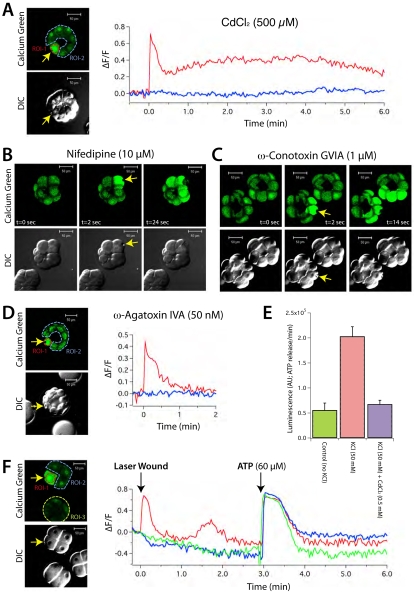
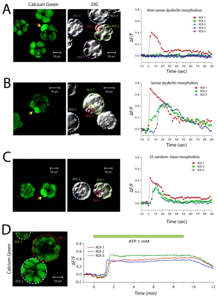
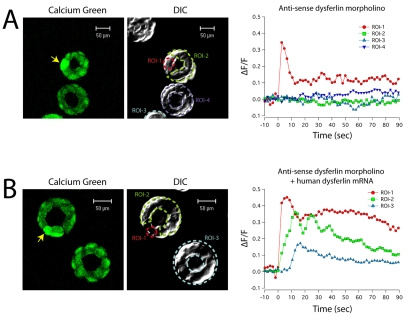
Dysferlin is a Ca(2+)-binding protein found in many different cell types. It is required for membrane wound repair in muscle, but it is not known whether it has the same function in other cells. Here we report the activation of an intercellular signaling pathway in sea urchin embryos by membrane wounding that evokes Ca(2+) spikes in neighboring cells. This pathway was mimicked by ATP application, and inhibited by apyrase, cadmium, and omega-agatoxin-IVA. Microinjection of dysferlin antisense phosphorodiamidate morpholino oligonucleotides blocked this pathway, whereas control morpholinos did not. Co-injection of mRNA encoding human dysferlin with the inhibitory morpholino rescued signaling activity. We conclude that in sea urchin embryos dysferlin mediates Ca(2+)-triggered intercellular signaling in response to membrane wounding.


Figures

Fig. 1.
Fig. 1.
Laser wounding triggers Ca2+ spikes in neighboring cells. (A) Differential interference contrast image (DIC, gray), FM 4-64 (red) and Calcium Green dextran (CGD) fluorescence (green) images of sea urchin embryos are shown 8 seconds prior to laser wounding (−8 sec), immediately after laser wounding a single blastomere (+2 sec), and at 14 (+14 sec) and 38 (+38 sec) seconds after laser wounding. Scale bars: 50 μm. (B) The timecourse of mean changes in CGD fluorescence (ΔF/F) are plotted for four ROIs (dashed red, blue, green and yellow regions in A): the blastomere wounded by the laser (red circles), two adjacent blastomeres that were not wounded (blue and green triangles), and the distal blastomere (yellow square).
Fig. 2.
Fig. 2.
Action at a distance. (A) Three non-contacting CGD-injected embryos were imaged by DIC and CGD two-photon microscopy immediately before (0 sec) and 4 (+4 sec) and 20 (+20 sec) seconds after laser wounding of a blastomere in one of the three embryos in the field (yellow arrow). (B) The timecourse of the CGD fluorescence change (ΔF/F) are plotted for four ROIs: the wounded blastomere (red circles), an adjacent blastomere in the wounded embryo (green open circles), and blastomeres in non-contacting embryos (blue and yellow triangles). ROIs 1-4 are demarcated on the DIC image in A by colored dashed circles.
Fig. 3.
Fig. 3.
Extracellular ATP triggers Ca2+ spikes in sea urchin embryos. (A) A CGD-injected embryo at the blastula stage was perfused with ASW supplemented with 1 mM ATP and imaged by time-lapse DIC and two-photon fluorescence microscopy. The timecourse of CGD fluorescence (ΔF/F) is plotted for four ROIs in four individual blastomeres in the same embryo. These ROIs are demarcated on the DIC image in A by red dashed circles. Green bars indicate the duration of ATP perfusion. Note the spontaneous non-synchronized Ca2+ spike activity observed in individual blastomeres prior to ATP perfusion (black arrows). (B) In Ca2+-free ASW (yellow bar) ATP perfusion (green bar) failed to trigger a rise in intracellular Ca2+ as measured by CGD fluorescence. Returning the embryo to ASW with normal Ca2+ (~9 mM; blue bar) restored the embryo's ability to respond to ATP perfusion. Note that in Ca2+-free ASW sea urchin tight junctions are destabilized with individual blastomeres assuming a rounded appearance in the DIC image. This rounded morphology did not immediately reverse upon perfusion with normal ASW. Despite this change in cell morphology, extracellular ATP could trigger intracellular Ca2+ spikes as long as extracellular Ca2+ was present.
Fig. 4.
Fig. 4.
Apyrase blocks the intercellular signaling triggered by laser wounding. CGD-injected embryo at the blastula stage was perfused with ASW supplemented with 1 mM ATP (A), ADP (B), or AMP (C) and imaged by time-lapse DIC and fluorescence microscopy. The timecourse of CGD fluorescence (ΔF/F) is plotted for ROIs encompassing the whole embryo, and are demarcated on the respective DIC image by red dashed circles. Green bars indicate the duration of nucleotide perfusion. (D) When a blastomere of a CGD-injected embryo was wounded with a laser (yellow arrow) in ASW supplemented with apyrase (20 IU/ml), Ca2+ spikes in adjacent blastomeres and in neighboring embryos were no longer observed.
Fig. 5.
Fig. 5.
Voltage-gated Ca2+ channels mediate the intercellular signaling triggered by wounding. A blastomere of a CGD-injected embryo was laser wounded (yellow arrow) in ASW supplemented with 500 μM CdCl2 (A), 10 μM nifedipine (B), 1 μM ω-conotoxin GVIA (C), or 50 nM ω-agatoxin IVA (D). (E) Depolarization of sea urchin embryos with 50 mM KCl triggered the release of ATP as measured using a firefly luciferase luminescence assay (compare green and red bars). ATP secretion in response to high KCl was blocked by 500 μM CdCl2 (blue bar). All points are mean ± s.d. n=19. (F) A blastomere of a CGD-injected embryo was laser wounded (yellow arrow) in ASW supplemented with 50 nM ω-agatoxin IVA. Three minutes after laser wounding, ATP (60 μM) was added to the bath and evoked Ca2+ spikes in the previously wounded blastomere (ROI-1, red line), in adjacent blastomeres in the same embryo (ROI-2, blue line), as well as in blastomeres in another CGD-injected embryo (ROI-3, green line).
Fig. 6.
Fig. 6.
Antisense dysferlin morpholinos inhibit intercellular signaling triggered by laser wounding. Sea urchin embryos were injected at the one-cell stage with CGD (5 mg/ml) and 1 mM antisense dysferlin morpholino (A), sense dysferlin morpholino (B), or a 25 random-base negative control morpholino (C). Following a 3-hour incubation period the embryos were imaged by time-lapse DIC and two-photon fluorescence microscopy before and after laser wounding a single blastomere (yellow arrow). The timecourse of mean CGD fluorescence changes (ΔF/F) is plotted for ROIs in the laser wounded blastomere (ROI-1, demarcated on the respective DIC image by red dashed circles; red symbols), blastomeres adjacent to the laser wounded blastomere (ROI-2, demarcated on the respective DIC image by a green dashed line; green symbols), and blastomeres in nearby non-contacting embryos. (ROIs 3-5, demarcated on the respective DIC image by blue and purple dashed lines, and correspondingly colored symbols). (D) Sea urchin embryos were injected at the one-cell stage with CGD and the antisense dysferlin morpholino. Injected embryos at the blastula stage were perfused with ASW supplemented with 1 mM ATP and imaged by time-lapse two-photon CGD fluorescence imaging. The timecourse of mean CGD fluorescence changes (ΔF/F) is plotted for three ROIs in the three different embryos. All three embryos responded to perfusion with ATP (green bar) with a rise in CGD fluorescence despite being injected with antisense dysferlin morpholino.
Fig. 7.
Fig. 7.
Co-injection of mRNA encoding human dysferlin rescues antisense dysferlin morpholino inhibition. (A) Sea urchin embryos were injected at the one-cell stage with CGD and the antisense dysferlin morpholino. Following a 3-hour incubation period the embryos were imaged by time-lapse DIC and two-photon fluorescence microscopy before and after laser wounding of a single blastomere (yellow arrow). The timecourse of mean CGD fluorescence changes (ΔF/F) is plotted for ROIs in the laser-wounded blastomere (ROI-1, demarcated on the respective DIC image by a red dashed line; red symbols), blastomeres adjacent to the laser-wounded blastomere (ROI-2, demarcated on the respective DIC image by green dashed line; green symbols), and blastomeres in nearby non-contacting embryos. (ROI-3 and 4, demarcated on the DIC image by blue and purple dashed lines, and correspondingly colored symbols). (B) Sea urchin embryos were injected at the one-cell stage with CGD, the antisense dysferlin morpholino, and mRNA encoding the 3HA-tagged human dysferlin. Following a 3-hour incubation period the embryos were imaged by time-lapse DIC and two-photon fluorescence microscopy before and after laser wounding of a single blastomere (yellow arrow). The timecourse of mean CGD fluorescence changes (ΔF/F) is plotted for ROIs in the laser-wounded blastomere (ROI-1, demarcated on the respective DIC image by red dashed lines; red symbols), blastomeres adjacent to the laser wounded blastomere (ROI-2, demarcated on the respective DIC image by a green dashed line; green symbols), and blastomeres in a nearby non-contacting embryo. (ROI-3, demarcated on the DIC image by a blue dashed line; blue symbols.)

References

    1. Angerer L. M., Angerer R. C. (2004). Disruption of gene function using antisense morpholinos. In Development of Sea Urchins, Ascidians, and other Invertebrate Deuterosomes: Experimental Approaches, vol. 74 (ed. Ettensohn C. A., Wessel G. M., Wray G. A.), p. 883 Amsterdam: Elsevier;
    1. Anselmi F., Hernandez V. H., Crispino G., Seydel A., Ortolano S., Roper S. D., Kessaris N., Richardson W., Rickheit G., Filippov M. A., et al. (2008). ATP release through connexin hemichannels and gap junction transfer of second messengers propagate Ca2+ signals across the inner ear. Proc. Natl. Acad. Sci. USA 105, 18770-18775 - PMC - PubMed
    1. Aoki M., Liu J., Richard I., Bashir R., Britton S., Keers S. M., Oeltjen J., Brown H. E., Marchand S., Bourg N., et al. (2001). Genomic organization of the dysferlin gene and novel mutations in Miyoshi myopathy. Neurology 57, 271-278 - PubMed
    1. Bansal D., Miyake K., Vogel S. S., Groh S., Chen C. C., Williamson R., McNeil P. L., Campbell K. P. (2003). Defective membrane repair in dysferlin-deficient muscular dystrophy. Nature 423, 168-172 - PubMed
    1. Bashir R., Strachan T., Keers S., Stephenson A., Mahjneh I., Marconi G., Nashef L., Bushby K. M. (1994). A gene for autosomal recessive limb-girdle muscular dystrophy maps to chromosome 2p. Hum. Mol. Genet. 3, 455-457 - PubMed

Publication types

MeSH terms