

Exercise training inhibits inflammatory cytokines and more than prevents myocardial dysfunction in rats with sustained beta-adrenergic hyperactivity
- PMID: 20442263
- PMCID: PMC2915518
- DOI: 10.1113/jphysiol.2010.187310
Exercise training inhibits inflammatory cytokines and more than prevents myocardial dysfunction in rats with sustained beta-adrenergic hyperactivity
Abstract
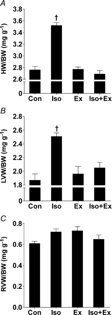
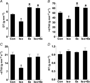
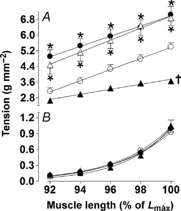
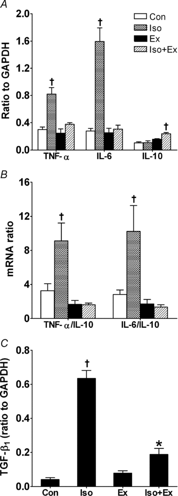
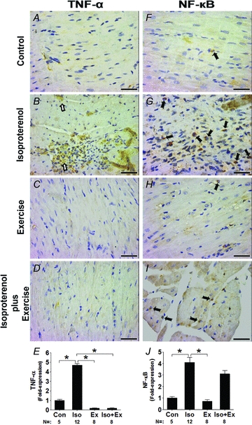
Myocardial hypertrophy and dysfunction occur in response to excessive catecholaminergic drive. Adverse cardiac remodelling is associated with activation of proinflammatory cytokines in the myocardium. To test the hypothesis that exercise training can prevent myocardial dysfunction and production of proinflammatory cytokines induced by beta-adrenergic hyperactivity, male Wistar rats were assigned to one of the following four groups: sedentary non-treated (Con); sedentary isoprenaline treated (Iso); exercised non-treated (Ex); and exercised plus isoprenaline (Iso+Ex). Echocardiography, haemodynamic measurements and isolated papillary muscle were used for functional evaluations. Real-time RT-PCR and Western blot were used to quantify tumour necrosis factor alpha, interleukin-6, interleukin-10 and transforming growth factor beta(1) (TGF-beta(1)) in the tissue. NF-B expression in the nucleus was evaluated by immunohistochemical staining. The Iso rats showed a concentric hypertrophy of the left ventricle (LV). These animals exhibited marked increases in LV end-diastolic pressure and impaired myocardial performance in vitro, with a reduction in the developed tension and maximal rate of tension increase and decrease, as well as worsened recruitment of the Frank-Starling mechanism. Both gene and protein levels of tumour necrosis factor alpha and interleukin-6, as well as TGF-beta(1) mRNA, were increased. In addition, the NF-B expression in the Iso group was significantly raised. In the Iso+Ex group, the exercise training had the following effects: (1) it prevented LV hypertrophy; (ii) it improved myocardial contractility; (3) it avoided the increase of proinflammatory cytokines and improved interleukin-10 levels; and (4) it attenuated the increase of TGF-beta(1) mRNA. Thus, exercise training in a model of beta-adrenergic hyperactivity can avoid the adverse remodelling of the LV and inhibit inflammatory cytokines. Moreover, the cardioprotection is related to beneficial effects on myocardial performance.
Figures

Comment in
-
Exercise training, inflammation and heart failure: working out to cool down.J Physiol. 2010 Jul 15;588(Pt 14):2525-6. doi: 10.1113/jphysiol.2010.194134. J Physiol. 2010. PMID: 20634182 Free PMC article. No abstract available.
Similar articles
-
Exercise promotes angiogenesis and improves beta-adrenergic receptor signalling in the post-ischaemic failing rat heart.Cardiovasc Res. 2008 May 1;78(2):385-94. doi: 10.1093/cvr/cvm109. Epub 2007 Dec 18. Cardiovasc Res. 2008. PMID: 18093988
-
Astragaloside IV attenuates inflammatory cytokines by inhibiting TLR4/NF-кB signaling pathway in isoproterenol-induced myocardial hypertrophy.J Ethnopharmacol. 2013 Dec 12;150(3):1062-70. J Ethnopharmacol. 2013. PMID: 24432369
-
Exercise training and beta-blocker treatment ameliorate age-dependent impairment of beta-adrenergic receptor signaling and enhance cardiac responsiveness to adrenergic stimulation.Am J Physiol Heart Circ Physiol. 2007 Sep;293(3):H1596-603. doi: 10.1152/ajpheart.00308.2007. Epub 2007 Jun 8. Am J Physiol Heart Circ Physiol. 2007. PMID: 17557919
-
The ageing spontaneously hypertensive rat as a model of the transition from stable compensated hypertrophy to heart failure.Eur Heart J. 1995 Dec;16 Suppl N:19-30. doi: 10.1093/eurheartj/16.suppl_n.19. Eur Heart J. 1995. PMID: 8682057 Review.
-
Cardiac hypertrophy induced by sustained beta-adrenoreceptor activation: pathophysiological aspects.Heart Fail Rev. 2007 Mar;12(1):66-86. doi: 10.1007/s10741-007-9007-4. Epub 2007 Mar 27. Heart Fail Rev. 2007. PMID: 17387610 Review.
Cited by
-
Xanthine Oxidase Inhibitor, Allopurinol, Prevented Oxidative Stress, Fibrosis, and Myocardial Damage in Isoproterenol Induced Aged Rats.Oxid Med Cell Longev. 2015;2015:478039. doi: 10.1155/2015/478039. Epub 2015 Jun 7. Oxid Med Cell Longev. 2015. PMID: 26137187 Free PMC article.
-
Exercise and Cardioprotection: A Natural Defense Against Lethal Myocardial Ischemia-Reperfusion Injury and Potential Guide to Cardiovascular Prophylaxis.J Cardiovasc Pharmacol Ther. 2019 Jan;24(1):18-30. doi: 10.1177/1074248418788575. Epub 2018 Jul 24. J Cardiovasc Pharmacol Ther. 2019. PMID: 30041547 Free PMC article. Review.
-
High-frequency ultrasound evaluation of effects of early treatment with metoprolol on myocardial inflammatory cytokine expression in rats with acute myocardial infarction.J Huazhong Univ Sci Technolog Med Sci. 2012 Oct;32(5):774-778. doi: 10.1007/s11596-012-1033-3. Epub 2012 Oct 18. J Huazhong Univ Sci Technolog Med Sci. 2012. PMID: 23073812
-
Treadmill exercise training prevents myocardial mechanical dysfunction induced by androgenic-anabolic steroid treatment in rats.PLoS One. 2014 Feb 12;9(2):e87106. doi: 10.1371/journal.pone.0087106. eCollection 2014. PLoS One. 2014. PMID: 24533053 Free PMC article.
-
Low-Level Laser Application in the Early Myocardial Infarction Stage Has No Beneficial Role in Heart Failure.Front Physiol. 2017 Jan 30;8:23. doi: 10.3389/fphys.2017.00023. eCollection 2017. Front Physiol. 2017. PMID: 28194115 Free PMC article.
References
-
- Barmeyer A, Müllerleile K, Mortensen K, Meinertz T. Diastolic dysfunction in exercise and its role for exercise capacity. Heart Fail Rev. 2009;14:125–134. - PubMed
-
- Boluyt MO, Long X, Eschenhagen T, Mende U, Schmitz W, Crow MT, Lakatta EG. Isoproterenol infusion induces alterations in expression of hypertrophy-associated genes in rat heart. Am J Physiol Heart Circ Physiol. 1995;269:H638–H647. - PubMed
-
- Brodowicz GR, Lamb DR. Exercise training, indomethacin, and isoproterenol induced myocardial necrosis in the rat. Basic Res Cardiol. 1991;86:40–48. - PubMed
Publication types
MeSH terms
Substances
LinkOut - more resources
Full Text Sources
Other Literature Sources
Medical