. 2010 Jul 1;588(Pt 13):2417-29.
doi: 10.1113/jphysiol.2010.188334. Epub 2010 May 4.

Cardiomyocytes with disrupted CFTR function require CaMKII and Ca(2+)-activated Cl(-) channel activity to maintain contraction rate

Affiliations

Cardiomyocytes with disrupted CFTR function require CaMKII and Ca(2+)-activated Cl(-) channel activity to maintain contraction rate

Zachary M Sellers et al. J Physiol. .

Abstract

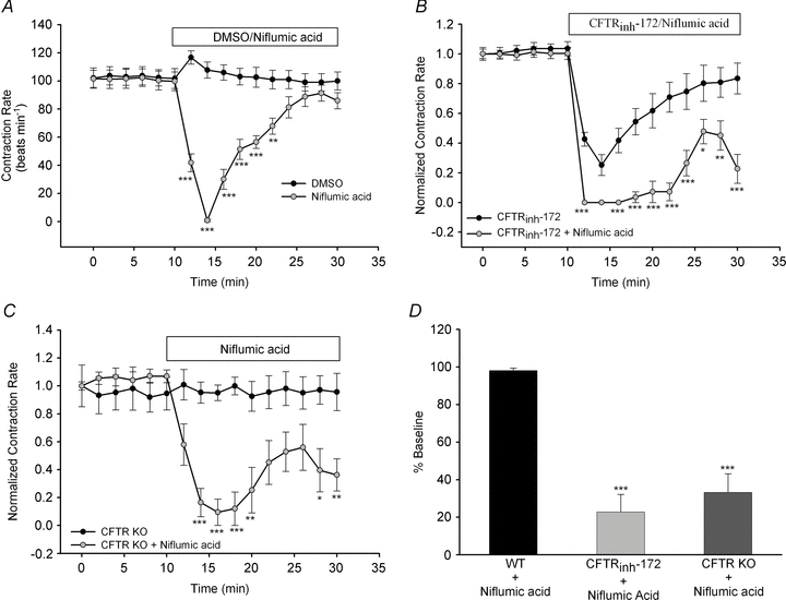
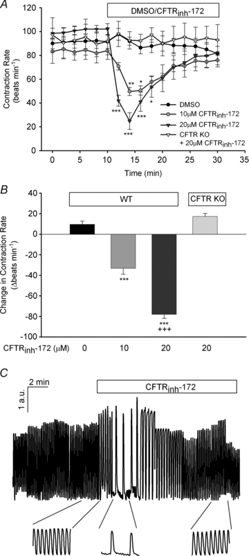
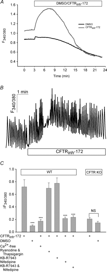
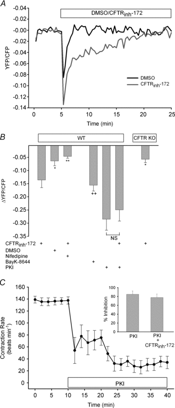
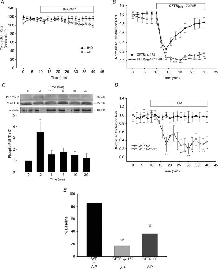
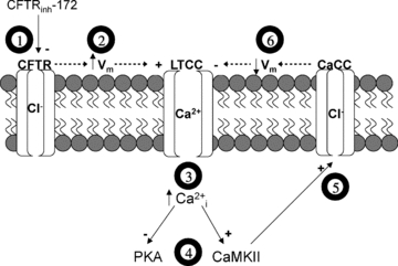
The physiological role of the cystic fibrosis transmembrane conductance regulator (CFTR) in cardiomyocytes remains unclear. Using spontaneously beating neonatal ventricular cardiomyocytes from wild-type (WT) or CFTR knockout (KO) mice, we examined the role of CFTR in the modulation of cardiomyocyte contraction rate. Contraction rates of spontaneously beating myocytes were captured by video imaging. Real-time changes in intracellular ([Ca(2+)](i)) and protein kinase A (PKA) activity were measured by fura-2 and fluorescence resonance energy transfer, respectively. Acute inhibition of CFTR in WT cardiomyocytes using the CFTR inhibitor CFTR(inh)-172 transiently inhibited the contraction rate. By contrast, cardiomyocytes from CFTR KO mice displayed normal contraction rates. Further investigation revealed that acute inhibition of CFTR activity in WT cardiomyocytes activated L-type Ca(2+) channels, leading to a transient increase of [Ca(2+)](i) and inhibition of PKA activity. Additionally, we found that contraction rate normalization following acute CFTR inhibition in WT cardiomyocytes or chronic deletion in cardiomyocytes from CFTR KO mice requires the activation of Ca(2+)/calmodulin-dependent kinase II (CaMKII) and Ca(2+)-activated Cl(-) channels (CaCC) because simultaneous addition of myristoylated-autocamtide-2-related inhibitory peptide or niflumic acid and CFTR(inh)-172 to WT cardiomyocytes or treatment of cardiomyoctes from CFTR KO mice with these agents caused sustained attenuation of contraction rates. Our results demonstrate that regulation of cardiomyocyte contraction involves CFTR. They also reveal that activation of CaMKII and CaCC compensates for loss of CFTR function. Increased dependence on CaMKII upon loss of CFTR function might leave cystic fibrosis patients at increased risk of heart dysfunction and disease.


Figures

Figure 5
Figure 5. Calcium-activated Cl channel plays an important role in cardiomyocyte contraction rate following CFTR inhibition or KO
A, syncytial WT ventricular myocytes were treated with DMSO (n= 6) or niflumic acid (100 μm; n= 7) for 30 min following a 10 min baseline period. B, separately, the effect of simultaneous addition of niflumic acid (100 μm) plus CFTRinh-172 (20 μm; n= 8) for 20 min was examined and compared with the effect of CFTRinh-172 alone (from Fig. 1A). C, ventricular myocytes from CFTR KO mice (n= 8) were treated with niflumic acid (100 μm) for 20 min after baseline measurements. Contraction rates were compared with those of the cardiomyocytes from the CFTR KO mice shown in Fig. 1A. *P < 0.05; **P < 0.01; ***P < 0.001 vs. cardiomyocytes from CFTR KO alone by two-way ANOVA. D, a comparison of changes in contraction rate due to niflumic acid in WT cardiomyocytes (n= 7), WT cardiomyocytes plus CFTRinh-172 (n= 8) and cardiomyocytes from CFTR KO mice (n= 8). ***P < 0.001 vs. WT by Student's unpaired t test.
Figure 1
Figure 1. Pharmacological inhibition, but not genetic deletion, of CFTR attenuates cardiomyocyte contraction rate
A, syncytial WT ventricular myocytes were treated with DMSO (n≥ 6) or CFTRinh-172 (10 μm or 20 μm; n= 7 each) for 20 min following a 10 min baseline period. Separately, the effect of CFTRinh-172 (20 μm; n= 5) on ventricular myocytes obtained from CFTR KO mice was also examined. *P < 0.05; **P < 0.01; ***P < 0.001 vs. DMSO or CFTR KO mice by two-way ANOVA. B, comparison of the maximal inhibitory effect of CFTRinh-172 on WT and CFTR KO cardiomyocyte contraction rate. ***P < 0.001 vs. DMSO by one-way ANOVA; +++P < 0.001 vs. 10 μm CFTRinh-172 by Student's unpaired t test. C, normalized representative trace of spontaneously beating WT ventricular myocyte with original traces prior to and after CFTRinh-172 (20 μm) addition. Deflection in the y-axis is expressed as arbitrary units (a.u.).
Figure 2
Figure 2. Inhibition of CFTR increases [Ca2+]i via L-type Ca2+ channels
A, quiescent WT ventricular myocytes were loaded with fura-2 to monitor changes in [Ca2+]i. Following a 3 min baseline period, either CFTRinh-172 (20 μm, n= 32) or DMSO (n= 19) was added, and subsequent effects were measured for 20 min. B, representative trace (n= 12) of spontaneously beating cardiomyocyte that was loaded with fura-2 to measure Ca2+ transients prior to and after CFTRinh-172 (20 μm) addition. Separate experiments with DMSO showed no change in diastolic or systolic Ca2+ (data not shown; n= 4). C, quiescent WT cardiomyocytes were bathed in Ca2+-free solution (n= 25), pretreated with ryanodine (10 μm) and thapsigargin (1 μm; n= 26), KB-R7943 (10 μm; n= 23), nifedipine (10 μm; n= 26) or KB-R7943 and nifedipine (n= 23) for 30 min prior to [Ca2+]i measurements. Additionally, in myocytes from CFTR KO mice, fura-2 fluorescence was monitored upon CFTRinh-172 (20 μm; n= 23) or DMSO (n= 24) addition. ***P < 0.001 vs. CFTRinh-172 by Student's unpaired t test; NS, not significant.
Figure 3
Figure 3. CFTRinh-172-induced activation of L-type Ca2+ channels causes inhibition of PKA and contraction rate
A, quiescent WT ventricular myocytes were infected with AKAR2.2 to monitor real-time changes in PKA activity. Following a 5 min baseline period, CFTRinh-172 (20 μm; n= 24) or DMSO (n= 18) was added, and subsequent effects were measured for 20 min. B, WT cardiomyocytes were pretreated with nifedipine (10 μm; n= 24) for 30 min prior to CFTRinh-172 addition. Additionally, PKA activity following CFTRinh-172 (20 μm; n= 19) was monitored in myocytes from CFTR KO mice. In separate experiments, the effects of BayK-8644 (1 μm; n= 15), PKI (20 μm; n= 14) or myristoylated PKI plus CFTRinh-172 (n= 14) on PKA activity in WT cardiomyocytes were measured. *P < 0.05; **P < 0.01 vs. CFTRinh-172; ++P < 0.01 vs. DMSO by Student's unpaired t test. C, syncytial WT ventricular myocytes were treated with PKI (20 μm; n= 8) for 30 min following a 10 min baseline period. Inset shows a comparison of syncytial WT cardiomyocytes treated with PKI alone or with PKI plus CFTRinh-172 (20 μm; n= 7).
Figure 4
Figure 4. Calcium/calmodulin-dependent kinase II activity is required to maintain normal contraction rates following pharmacological inhibition or genetic deletion of CFTR
A, syncytial WT ventricular myocytes were treated with H2O (n= 5) or AIP (1 μm, n= 6) for 30 min following a 10 min baseline period. B, separately, the effect of simultaneous addition of AIP plus CFTRinh-172 (20 μm; n= 8) for 20 min was examined and compared with responses by CFTRinh-172 alone (from Fig. 1A). *P < 0.05; ***P < 0.001 vs. CFTRinh-172 alone by two-way ANOVA. C, WT cardiomyocytes were exposed to CFTRinh-172 (20 μm) for various lengths of time (n= 3–5 each) and then harvested to examine PLB-Thr17 phosphorylation. All results were quantified and normalized. A representative blot is shown. *P < 0.05 vs. no treatment by Student's unpaired t test. D, cardiomyocytes from CFTR KO mice (n= 8) were treated with AIP (1 μm) for 30 min following baseline measurements. Contraction rates were compared with those of cardiomyocytes from CFTR KO mice shown in Fig. 1A. *P < 0.05; **P < 0.01 vs. cardiomyocytes from CFTR KO alone by two-way ANOVA. E, a comparison of changes in contraction rate due to AIP in WT cardiomyocytes (n= 6), WT cardiomyocytes plus CFTRinh-172 (n= 8) and cardiomyocytes from CFTR KO mice (n= 8). **P < 0.01; ***P < 0.001 vs. WT by Student's unpaired t test.
Figure 6
Figure 6. Proposed model for the effect of CFTR disruption on cardiomyocyte signalling and ion transport
Acute inhibition of CFTR by CFTRinh-172 (1) probably causes an increase in membrane potential (Vm), which leads to increased activation of L-type Ca2+ channels (LTCC) (2). Increased LTCC activation raises intracellular Ca2+ (3), resulting in inhibition of PKA and activation of CaMKII (4). Inhibition of PKA causes a decrease in contraction rate. Activation of CaMKII stimulates Ca2+-activated Cl channels (CaCC) (5), which probably serves to restore changes in membrane potential and decrease LTCC hyperactivity (6). This decrease in [Ca2+]i probably removes the inhibition on PKA activity, thus restoring contraction rate.

References

    1. Ambrosi P, Chazalettes JP, Viard L, Raynaud M, Faugere G, Noirclerc M, Bernard PJ. Left ventricular involvement in mucoviscidosis after 2 years of age. Arch Fr Pediatr. 1993;50:653–656. - PubMed
    1. Anderson ME. Multiple downstream proarrhythmic targets for calmodulin kinase II: moving beyond an ion channel-centric focus. Cardiovasc Res. 2007;73:657–666. - PubMed
    1. Bers DM. Calcium cycling and signalling in cardiac myocytes. Annu Rev Physiol. 2008;70:23–49. - PubMed
    1. Berul CI, Sweeten T, Vetter VL, Morad M. Lack of cystic fibrosis transmembrane regulator-type chloride current in pediatric human atrial myocytes. Life Sci. 1997;60:189–197. - PubMed
    1. Chen H, Liu LL, Ye LL, McGuckin C, Tamowski S, Scowen P, Tian H, Murray K, Hatton WJ, Duan D. Targeted inactivation of cystic fibrosis transmembrane conductance regulator chloride channel gene prevents ischemic preconditioning in isolated mouse heart. Circulation. 2004;110:700–704. - PubMed

Publication types

MeSH terms

Substances

LinkOut - more resources