

Two closely related genes of Arabidopsis encode plastidial cytidinediphosphate diacylglycerol synthases essential for photoautotrophic growth
- PMID: 20442275
- PMCID: PMC2899908
- DOI: 10.1104/pp.110.156422
Two closely related genes of Arabidopsis encode plastidial cytidinediphosphate diacylglycerol synthases essential for photoautotrophic growth
Abstract
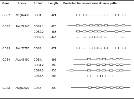
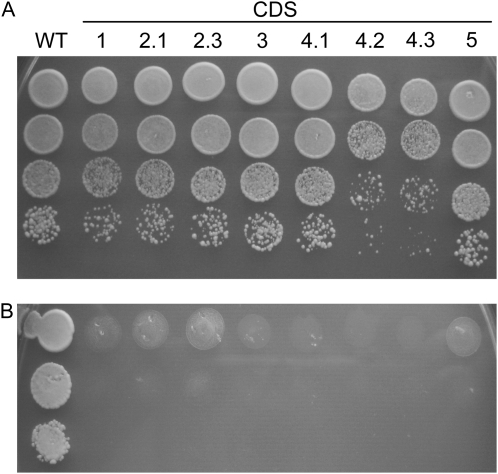
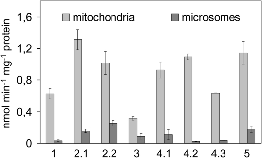
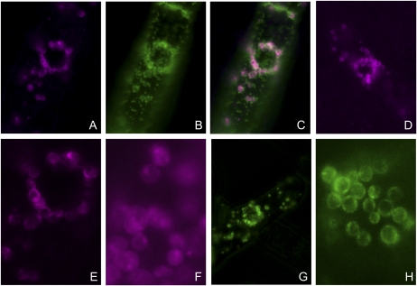
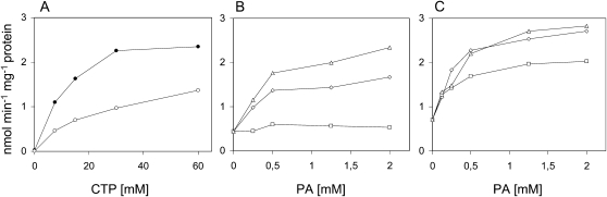
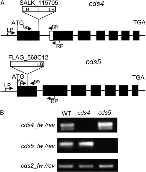
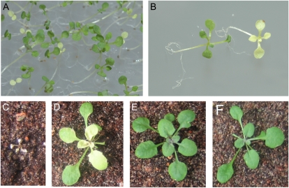
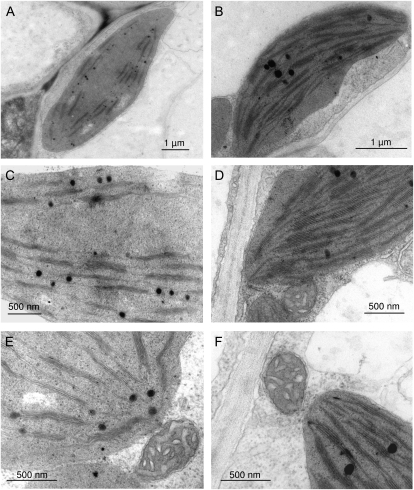
Cytidinediphosphate diacylglycerol synthase (CDS) catalyzes the formation of cytidinediphosphate diacylglycerol, an essential precursor of anionic phosphoglycerolipids like phosphatidylglycerol or -inositol. In plant cells, CDS isozymes are located in plastids, mitochondria, and microsomes. Here, we show that these isozymes are encoded by five genes in Arabidopsis (Arabidopsis thaliana). Alternative translation initiation or alternative splicing of CDS2 and CDS4 transcripts can result in up to 10 isoforms. Most of the cDNAs encoding the various plant isoforms were functionally expressed in yeast and rescued the nonviable phenotype of the mutant strain lacking CDS activity. The closely related genes CDS4 and CDS5 were found to encode plastidial isozymes with similar catalytic properties. Inactivation of both genes was required to obtain Arabidopsis mutant lines with a visible phenotype, suggesting that the genes have redundant functions. Analysis of these Arabidopsis mutants provided further independent evidence for the importance of plastidial phosphatidylglycerol for structure and function of thylakoid membranes and, hence, for photoautotrophic growth.
Figures
Similar articles
-
Extraplastidial cytidinediphosphate diacylglycerol synthase activity is required for vegetative development in Arabidopsis thaliana.Plant J. 2013 Sep;75(5):867-79. doi: 10.1111/tpj.12248. Epub 2013 Jun 21. Plant J. 2013. PMID: 23711240
-
Plastidial glycolysis in developing Arabidopsis embryos.New Phytol. 2010 Feb;185(3):649-62. doi: 10.1111/j.1469-8137.2009.03113.x. Epub 2009 Dec 9. New Phytol. 2010. PMID: 20002588
-
Cytidine diphosphate diacylglycerol synthase is essential for mitochondrial structure and energy production in Arabidopsis thaliana.Plant J. 2023 Apr;114(2):338-354. doi: 10.1111/tpj.16139. Epub 2023 Feb 28. Plant J. 2023. PMID: 36789486
-
Plastidial glyceraldehyde-3-phosphate dehydrogenase deficiency leads to altered root development and affects the sugar and amino acid balance in Arabidopsis.Plant Physiol. 2009 Oct;151(2):541-58. doi: 10.1104/pp.109.143701. Epub 2009 Aug 12. Plant Physiol. 2009. PMID: 19675149 Free PMC article.
-
CDP-diacylglycerol synthase of microorganisms.Biochim Biophys Acta. 1997 Sep 4;1348(1-2):157-65. doi: 10.1016/s0005-2760(97)00111-2. Biochim Biophys Acta. 1997. PMID: 9370328 Review.
Cited by
-
Arabidopsis Sec14 proteins (SFH5 and SFH7) mediate interorganelle transport of phosphatidic acid and regulate chloroplast development.Proc Natl Acad Sci U S A. 2023 Feb 7;120(6):e2221637120. doi: 10.1073/pnas.2221637120. Epub 2023 Jan 30. Proc Natl Acad Sci U S A. 2023. PMID: 36716376 Free PMC article.
-
Acyl-lipid metabolism.Arabidopsis Book. 2013;11:e0161. doi: 10.1199/tab.0161. Epub 2013 Jan 29. Arabidopsis Book. 2013. PMID: 23505340 Free PMC article.
-
Lipidomics-Assisted GWAS (lGWAS) Approach for Improving High-Temperature Stress Tolerance of Crops.Int J Mol Sci. 2022 Aug 20;23(16):9389. doi: 10.3390/ijms23169389. Int J Mol Sci. 2022. PMID: 36012660 Free PMC article. Review.
-
Mitochondrial CDP-diacylglycerol synthase activity is due to the peripheral protein, TAMM41 and not due to the integral membrane protein, CDP-diacylglycerol synthase 1.Biochim Biophys Acta Mol Cell Biol Lipids. 2018 Mar;1863(3):284-298. doi: 10.1016/j.bbalip.2017.12.005. Epub 2017 Dec 16. Biochim Biophys Acta Mol Cell Biol Lipids. 2018. PMID: 29253589 Free PMC article.
-
Arabidopsis ACYL CARRIER PROTEIN4 and RHOMBOID LIKE10 act independently in chloroplast phosphatidate synthesis.Plant Physiol. 2023 Nov 22;193(4):2661-2676. doi: 10.1093/plphys/kiad483. Plant Physiol. 2023. PMID: 37658850 Free PMC article.
References
-
- Andrews J, Mudd JB. (1984) Characterization of CDP-DG and PG synthesis in pea chloroplasts. Siegenthaler PA, Eichenberger W, , Structure, Function and Metabolism of Plant Lipids. Elsevier North Holland Biomedical Press, Amsterdam, pp 131–134
-
- Babiychuk E, Müller F, Eubel H, Braun HP, Frentzen M, Kushnir S. (2003) Arabidopsis phosphatidylglycerophosphate synthase 1 is essential for chloroplast differentiation, but is dispensable for mitochondrial function. Plant J 33: 899–909 - PubMed
-
- Bechtold N, Ellis J, Pelletier G. (1993) In planta Agrobacterium mediated gene transfer by infiltration of adult Arabidopsis thaliana plants. C R Acad Sci III 316: 1194–1199
Publication types
MeSH terms
Substances
LinkOut - more resources
Full Text Sources
Molecular Biology Databases
Research Materials
