. 2010 Jul;299(1):E23-32.
doi: 10.1152/ajpendo.00664.2009. Epub 2010 May 4.

An AP-3-dependent mechanism drives synaptic-like microvesicle biogenesis in pancreatic islet beta-cells

Affiliations

An AP-3-dependent mechanism drives synaptic-like microvesicle biogenesis in pancreatic islet beta-cells

Arthur T Suckow et al. Am J Physiol Endocrinol Metab. 2010 Jul.

Abstract

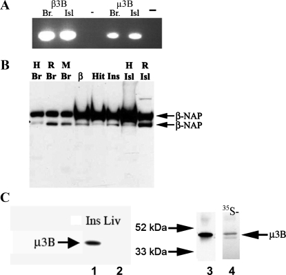
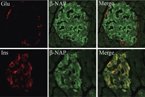
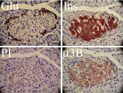
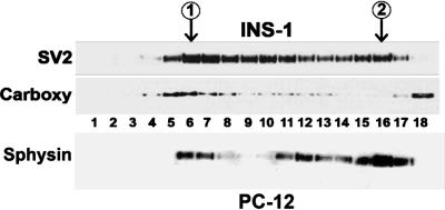
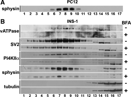
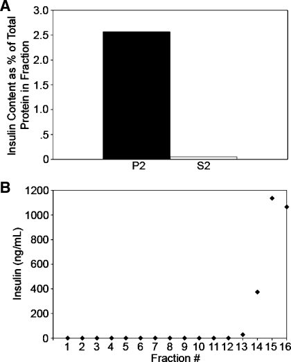
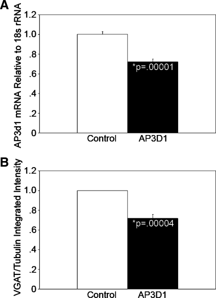
Pancreatic islet beta-cells contain synaptic-like microvesicles (SLMVs). The origin, trafficking, and role of these SLMVs are poorly understood. In neurons, synaptic vesicle (SV) biogenesis is mediated by two different cytosolic adaptor protein complexes, a ubiquitous AP-2 complex and the neuron-specific AP-3B complex. Mice lacking AP-3B subunits exhibit impaired GABAergic (inhibitory) neurotransmission and reduced neuronal vesicular GABA transporter (VGAT) content. Since beta-cell maturation and exocytotic function seem to parallel that of the inhibitory synapse, we predicted that AP-3B-associated vesicles would be present in beta-cells. Here, we test the hypothesis that AP-3B is expressed in islets and mediates beta-cell SLMV biogenesis. A secondary aim was to test whether the sedimentation properties of INS-1 beta-cell microvesicles are identical to those of bona fide SLMVs isolated from PC12 cells. Our results show that the two neuron-specific AP-3 subunits beta3B and mu3B are expressed in beta-cells, the first time these proteins have been found to be expressed outside the nervous system. We found that beta-cell SLMVs share the same sedimentation properties as PC12 SLMVs and contain SV proteins that sort specifically to AP-3B-associated vesicles in the brain. Brefeldin A, a drug that interferes with AP-3-mediated SV biogenesis, inhibits the delivery of AP-3 cargoes to beta-cell SLMVs. Consistent with a role for AP-3 in the biogenesis of GABAergic SLMV in beta-cells, INS-1 cell VGAT content decreases upon inhibition of AP-3 delta-subunit expression. Our findings suggest that beta-cells and neurons share molecules and mechanisms important for mediating the neuron-specific membrane trafficking pathways that underlie synaptic vesicle formation.


Figures

Fig. 1.
Fig. 1.
Expression of neuronal adaptor protein (AP)-3 mRNA and protein in INS-1 β-cells and pancreatic islet extracts. A: PCR analysis was performed to determine whether the neuronal AP-3 subunits β3B and μ3B are expressed in human islets (Isl). Human brain (Br) cDNA was used as a positive control. PCR products of the expected size were detected in brain and islets for both β3B and μ3B. A negative control (−) in which no reverse transcriptase was added during the reverse transcription reaction (no RT control) is shown to the right of the brain and islet lanes. cDNA from human tissues (shown) and from rat tissues (not shown) yielded identical results. B: to determine whether β3B protein is expressed in islets, Western blot analysis was performed using a β3B-specific monoclonal antibody. As expected, β3B was detected in human (H), rat (R), and mouse (M) Br. β3B protein was also present in cell lysates from the insulin-secreting β-cell lines β-TC3 (β), HIT-T15 (Hit), and INS-1 (Ins) and in tissue lysates from human and rat Isl. β3B was not detected in human liver or in brain from β3B-knockout mice (Ref. and data not shown). As observed previously, immunoblot detection of β3B yielded 2 bands (Mr of ∼140 K). C, left, lanes 1 and 2: μ3B was detected in INS-1 cell lysate (Ins) but not in rat liver lysate (Liv). C, right: μ3B produced by in vitro transcription and translation (lane 4) and analyzed by immunoblotting with the μ3B antibody ran at the expected Mr of ∼47,000. This band comigrated precisely with the band that was observed in INS-1 cells (lane 3). 35S-met was included in the in vitro translation reaction. Autoradiographic detection of the radiolabeled μ3B (lane 4) yielded a band that precisely aligned with the band detected by immunoblot analysis. This confirms that the μ3B antibody detected μ3B synthesized de novo in the in vitro transcription and translation reaction.
Fig. 2.
Fig. 2.
β3B is detected in the pancreatic islet β-cells. Immunofluorescent localization was performed to determine the localization of β3B (β-NAP; green) in rat pancreas sections. Tissues were costained for either insulin (Ins; bottom, red) or glucagon (Glu; top, red). β3B was detected in cells expressing insulin (colocalization yields orange-yellow in the merged images) but not in cells expressing glucagon. As expected, β3B was also not present in the surrounding pancreatic exocrine tissue. Islet-specific expression of β3B was also observed by immunostaining using the same antibody in mouse and human pancreas sections (not shown). Magnification, ×60.
Fig. 3.
Fig. 3.
Localization of μ3B in pancreatic islets. Immunohistochemical localization was performed to determine the localization of μ3B in rat pancreas sections. Serial sections were stained for either Ins, Glu, or, as a control, preimmune serum (PI). Four serial sections containing the same representative islet are shown. As is typical in rodent islets, the glucagon-expressing α-cells are found in the islet periphery, surrounding the insulin-producing β-cells. μ3B was observed in the β-cells but not in cells that stained positive for glucagon. As expected, μ3B was also not present in the surrounding pancreatic exocrine tissue. PI yielded very little background. Magnification, ×60.
Fig. 4.
Fig. 4.
Detection of synaptic-like microvesicles (SLMVs) in INS-1 cell homogenates by sucrose gradient sedimentation. Postnuclear supernatant (S1) from INS-1 cells was sedimented in 5–45% sucrose gradients, and the resulting fractions were analyzed by immunoblotting. When fractions were stained with synaptic vesicle-2 (SV2) antibody, 2 distinct peaks [labeled peak 1 and peak 2 (inside circles)] were observed at fractions 6 and 16. Larger vesicles sediment to the lower-numbered fractions, and peak 1 results from the presence of SV2 in larger endosomes and secretory granules. The presence of secretory granule proteins in peak 1 was confirmed by immunoblotting for a specific marker of the secretory granules, carboxypeptidase E (carboxy). The presence of a 2nd peak centered at fraction 16 is consistent with the presence of SLMVs in INS-1 cells and with the association of SV2 with the INS-1 cell SLMVs. The absence of carboxy from the 2nd peak demonstrates that the population of microvesicles comprising peak 2 was not contaminated with secretory granules. Carboxy released from broken secretory granules was, as expected, present in the pool of soluble proteins in fraction 18. An immunoblot from PC12 cells fractionated in parallel (bottom row) was probed for synaptophysin (sphysin), which localizes primarily to SLMV. Sphysin content formed a peak around fraction 16, demonstrating that the pattern of PC12 SLMV sedimentation in the sucrose gradient matches that of INS-1 SLMV. Lesser amounts of sphysin are also present in larger PC12 cell compartments, including secretory granules, which explains the presence of bands in PC12 fractions 6 and 7 (40, 41).
Fig. 5.
Fig. 5.
SLMV biogenesis in INS-1 β-cells is sensitive to brefeldin A (BFA) inhibition. PC12 cells produce SLMVs with biophysical properties indistinguishable from those of neuronal SVs. A: high-speed supernatants (S2) obtained from PC12 cells were fractionated by glycerol gradient centrifugation, a technique especially well suited for resolving the ∼40-nm SVs and SLMVs. Fractions containing SLMVs were identified using an antibody to the SV marker sphysin, a known PC12 SLMV protein. Maximal immunoreactivity to sphysin was observed in fraction 8. B: high-speed S2 supernatants obtained from INS-1 cells that had been incubated for 2 h in the presence (+ sign in column on the far right) or absence (−) of BFA were sedimented in 5–25% glycerol velocity gradients to resolve SLMV. Fractions (numbered 1–17) were analyzed by immunoblotting using antibodies to proteins known to be associated with SVs and PC12 SLMVs. As in PC12 cells, which were analyzed in parallel, maximal immunoreactivity to the SV marker sphysin peaked at fraction 8, demonstrating that the SLMVs in the 2 cell types are the same size. All other SLMV markers tested also peaked at fraction 8, including phosphatidylinositol 4-kinase type IIα (PI4KIIα), an SV protein sorted specifically by an AP-3B-dependent mechanism. Treatment with BFA markedly reduced the content of vacuolar ATPase (vATPase), SV2, PI4KIIα, and sphysin in the INS-1 SLMV fractions (compare fractions 6–9 in the + rows with the same fractions in the − rows directly above). The observed depletion of protein content in the INS-1 SLMV fractions during BFA treatment is identical to previous results demonstrating AP-3-mediated SLMV biogenesis using BFA in PC12 cells (18, 37, 38). Probing for tubulin (bottom 2 rows, fractions 14–17) confirmed that the glycerol gradients were loaded with extracts with equal protein content. Cytosolic proteins like tubulin are expected to localize to the upper fractions, as is seen here. + and − blots were run, transferred, and exposed to film for equal lengths of time in parallel.
Fig. 6.
Fig. 6.
SLMVs do not contain detectable amounts of insulin. To confirm that the analyzed vesicles identified as being SLMV were devoid of insulin-containing secretory granules, the insulin content of the crude SLMV-containing S2 fraction was compared with the secretory granule-containing P2 fraction. A: bars represent insulin content as %total protein content in the fraction; 0.06% of the total protein in the S2 fraction represented insulin, whereas 2.57% percent of the protein in the P2 fraction represented insulin. B: the sedimentation of residual insulin in the S2 fraction was determined by glycerol velocity sedimentation and analysis of the resulting fractions for insulin content; the concentration of insulin in each fraction is shown. Insulin was detected in the cytosolic fractions where tubulin is found (fractions 13 and above) and was absent from all of the fractions containing the SLMVs (fractions 3–12).
Fig. 7.
Fig. 7.
Knockdown of the AP-3 δ-subunit reduces the expression of the GABAergic SV marker vesicular GABA transporter (VGAT). A: INS-1 cells were transduced with lentiviral particles expressing short hairpin (sh)RNA against the AP-3δ1 subunit of the AP-3 complex or with control nontargeting shRNA. After selection for cells stably expressing the viral constructs, knockdown was confirmed by quantitative RT-PCR. AP-3δ1 mRNA levels are shown relative to the level in cells expressing the control shRNA and were 28% less in AP-3δ1 shRNA-expressing INS-1 cells (right) than in INS-1 cells stably expressing the nontargeting shRNA (left; n = 3, P < 0.00002). B: quantitative Western blot analysis of protein lysates revealed a 28% reduction in VGAT content in AP-3δ1 shRNA-expressing INS-1 cells (right) relative to nontargeting shRNA control INS-1 cells (left; n = 5, P < 0.00004). Five separate control and AP-3-knockdown lysate samples with equal protein content were analyzed in duplicate or triplicate on separate immunoblots. VGAT and tubulin signal intensities were measured using an Odyssey infrared imaging system. VGAT signal intensities were normalized to tubulin. The anti-VGAT antibody has previously been characterized by us in detail (9, 47).

Similar articles

Cited by

References

    1. Abderrahmani A, Niederhauser G, Plaisance V, Haefliger JA, Regazzi R, Waeber G. Neuronal traits are required for glucose-induced insulin secretion. FEBS Lett 565: 133–138, 2004 - PubMed
    1. Blumstein J, Faundez V, Nakatsu F, Saito T, Ohno H, Kelly RB. The neuronal form of adaptor protein-3 is required for synaptic vesicle formation from endosomes. J Neurosci 21: 8034–8042, 2001 - PMC - PubMed
    1. Boehm M, Bonifacino JS. Adaptins: the final recount. Mol Biol Cell 12: 2907–2920, 2001 - PMC - PubMed
    1. Bonifacio E, Lernmark A, Dawkins RL. Serum exchange and use of dilutions have improved precision of measurement of islet cell antibodies. J Immunol Methods 106: 83–88, 1988 - PubMed
    1. Braun M, Wendt A, Birnir B, Broman J, Eliasson L, Galvanovskis J, Gromada J, Mulder H, Rorsman P. Regulated exocytosis of GABA-containing synaptic-like microvesicles in pancreatic beta-cells. J Gen Physiol 123: 191–204, 2004 - PMC - PubMed

Publication types

MeSH terms