Comparative Study
. 2010 May 18;107(20):9222-7.
doi: 10.1073/pnas.1004584107. Epub 2010 May 4.

Human embryonic stem cells with biological and epigenetic characteristics similar to those of mouse ESCs

Affiliations
Comparative Study

Human embryonic stem cells with biological and epigenetic characteristics similar to those of mouse ESCs

Jacob Hanna et al. Proc Natl Acad Sci U S A. .

Abstract

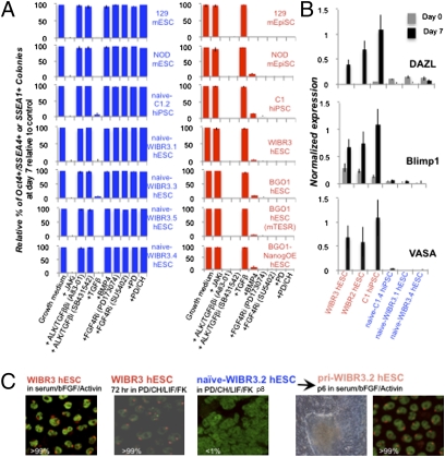
Human and mouse embryonic stem cells (ESCs) are derived from blastocyst-stage embryos but have very different biological properties, and molecular analyses suggest that the pluripotent state of human ESCs isolated so far corresponds to that of mouse-derived epiblast stem cells (EpiSCs). Here we rewire the identity of conventional human ESCs into a more immature state that extensively shares defining features with pluripotent mouse ESCs. This was achieved by ectopic induction of Oct4, Klf4, and Klf2 factors combined with LIF and inhibitors of glycogen synthase kinase 3beta (GSK3beta) and mitogen-activated protein kinase (ERK1/2) pathway. Forskolin, a protein kinase A pathway agonist which can induce Klf4 and Klf2 expression, transiently substitutes for the requirement for ectopic transgene expression. In contrast to conventional human ESCs, these epigenetically converted cells have growth properties, an X-chromosome activation state (XaXa), a gene expression profile, and a signaling pathway dependence that are highly similar to those of mouse ESCs. Finally, the same growth conditions allow the derivation of human induced pluripotent stem (iPS) cells with similar properties as mouse iPS cells. The generation of validated "naïve" human ESCs will allow the molecular dissection of a previously undefined pluripotent state in humans and may open up new opportunities for patient-specific, disease-relevant research.


Conflict of interest statement

Conflict of interest statement: R.J. is a cofounder of Fate Therapeutics and an adviser to Stemgent. R.J. and J.H. have filed a patent application describing the results and concepts presented herein.

Figures

Fig. 1.
Fig. 1.
Derivation of naïve mouse ESC-like induced pluripotent stem cells. (A) Strategy and representative images of C1 cultures and subcloned cell line C1.2 observed at different stages during reprogramming. p, passage number. NOD mESCs and C1.2 hiPSCs after DOX withdrawal are also shown. (B) C1 hiPSC line maintained in conventional bFGF/serum-supplemented human ES growth conditions (hESM) was transferred into N2B27 PD/CH/LIF + DOX, and emerging colonies were subcloned. Representative C1.10 hiPSC clone is shown. (C) Signaling dependence of pluripotent cell lines. Pluripotent cells were equally divided and plated on feeders in the indicated growth medium in which these cell lines are normally maintained, and 36 h later the wells were supplemented with the indicated inhibitors or growth factors. After 6 days, wells were fixed and stained for Nanog to determine the relative percentage of pluripotent colonies. Colony formation is normalized to an internal control growth medium only without inhibitors. (D) C1.2 hiPSC line was electroporated with mammalian expression vectors expressing the indicated reprogramming factors and cells were subjected to puromycin selection and passaged in PD/CH/LIF without DOX. Values indicate relative percentage of SSEA4+ colonies obtained in comparison with control cells that were transfected with an Oct4/Klf4/Sox2-encoding polycistronic construct. (E) Screening of factors that allow propagation of transgene-independent (i.e., DOX-independent) C1 hiPSCs in PD/CH/LIF-supplemented media. Effect of the removal of individual factors from the pool of 13 indicated small molecules or cytokines on the stabilization of pluripotent C1 hiPSCs independent of DOX. C1 cells were plated on feeders in N2B27 media with the indicated factors. P values using Student's t test indicate significant change in comparison with cells grown in DOX/PD/CH/LIF conditions, which were defined as a control (100% survival).
Fig. 2.
Fig. 2.
Characteristics of naïve hESC lines. (A) Scheme for reverting hESCs to generate naïve hESCs. Representative images of WIBR3 hESCs at different stages of the reversion process in PD/CH/LIF/FK. p, passage number. Magnifications of captured images are indicated. (B) Single-cell cloning efficiency of different pluripotent stem cell lines as determined by the number of wells containing Nanog+ colonies after 7 days. (C) Estimated cell doubling time. Plated cells were plated in triplicates and counted at 1, 4, and 7 days after plating, and increase in cell number was used to extrapolate average doubling time. Error bars represent SD, and P values using Student's t test indicate significant difference in the average of hESC/hiPSC lines in comparison with the average of naïve hESC/hiPSC lines.
Fig. 3.
Fig. 3.
Naïve hESCs share defining signaling and epigenetic features with mESCs. (A) Signaling dependence of pluripotent cell lines conducted as in Fig. 1C. After 7 days, wells were fixed and stained to determine the relative percentage of colonies positive for pluripotency markers. SSEA1 staining was used for mouse stem cells. Colony formation was normalized to an internal control growth medium without inhibitors (first left column). Normalized percentages lower than 5% are defined as “sensitivity” to the presence of the supplemented inhibitor. (B) RT-PCR expression of early germ-cell markers in the presence or absence of BMP4/7/8 cytokines. (C) Representative fluorescence in situ hybridization (FISH) analysis for XIST RNA (red) and Cot1 nuclear RNA (green). Pri-WIBR3.2 cell line was analyzed after passaging in conventional bFGF/serum-containing human ESC growth conditions. Numbers indicate average percentage of XIST-positive nuclei counted.
Fig. 4.
Fig. 4.
Naïve hESCs/hiPSCs share a global transcriptional profile with mESCs. (A) Bar chart showing median expression ratio of pluripotency and lineage-specific marker genes in hESCs and naïve hESCs. Asterisks delineate genes in which the false discovery rate was <0.1 between the naïve and primed group of samples. (B) FACS analysis for surface expression of human and mouse MHC class I alleles. Black graph indicates isotype match control. MFI, median fluorescence intensity. P values using Student's t test indicate significant change (P < 0.01). (C) Cross-species gene expression clustering where mESCs and naïve hESCs formed a distinct group apart from mEpiSCs and hESCs. Legend shown on right with yellow and blue indicating positive and negative correlation, respectively. Gene expression relative abundance was clustered by Spearman correlation and average linkage. Mouse samples are labeled in purple and human samples are labeled in brown.
Fig. 5.
Fig. 5.
Metastable states of pluripotency. Model describing relationships between genetic background and requirements for exogenous factors to achieve in vitro stabilization of the naïve (ICM-like or ESC-like) and primed (epiblast-like or EpiSC-like) pluripotent states. “Metastability” pertains to describing a system with two or more in vitro stable states that can interconvert by defined signals. The transcription factors and culture supplements minimally required for interconversion and stabilization of the respective pluripotent states in the different strains and species are highlighted.

References

    1. Evans MJ, Kaufman MH. Establishment in culture of pluripotential cells from mouse embryos. Nature. 1981;292:154–156. - PubMed
    1. Jaenisch R, Young R. Stem cells, the molecular circuitry of pluripotency and nuclear reprogramming. Cell. 2008;132:567–582. - PMC - PubMed
    1. Tesar PJ, et al. New cell lines from mouse epiblast share defining features with human embryonic stem cells. Nature. 2007;448:196–199. - PubMed
    1. Brons IG, et al. Derivation of pluripotent epiblast stem cells from mammalian embryos. Nature. 2007;448:191–195. - PubMed
    1. Nichols J, Smith A. Naive and primed pluripotent states. Cell Stem Cell. 2009;4:487–492. - PubMed

Publication types

MeSH terms