. 2010 May;22(5):1592-604.
doi: 10.1105/tpc.109.073577. Epub 2010 May 4.

Alteration of substrate specificity: the variable N-terminal domain of tobacco Ca(2+)-dependent protein kinase is important for substrate recognition

Affiliations

Alteration of substrate specificity: the variable N-terminal domain of tobacco Ca(2+)-dependent protein kinase is important for substrate recognition

Takeshi Ito et al. Plant Cell. 2010 May.

Abstract

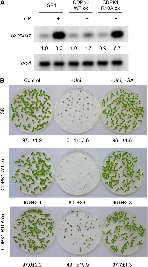
Protein kinases are major signaling molecules that are involved in a variety of cellular processes. However, the molecular mechanisms whereby protein kinases discriminate specific substrates are still largely unknown. Ca(2+)-dependent protein kinases (CDPKs) play central roles in Ca(2+) signaling in plants. Previously, we found that a tobacco (Nicotiana tabacum) CDPK1 negatively regulated the transcription factor REPRESSION OF SHOOT GROWTH (RSG), which is involved in gibberellin feedback regulation. Here, we found that the variable N-terminal domain of CDPK1 is necessary for the recognition of RSG. A mutation (R10A) in the variable N-terminal domain of CDPK1 reduced both RSG binding and RSG phosphorylation while leaving kinase activity intact. Furthermore, the R10A mutation suppressed the in vivo function of CDPK1. The substitution of the variable N-terminal domain of an Arabidopsis thaliana CDPK, At CPK9, with that of Nt CDPK1 conferred RSG kinase activities. This chimeric CDPK behaved according to the identity of the variable N-terminal domain in transgenic plants. Our results open the possibility of engineering the substrate specificity of CDPK by manipulation of the variable N-terminal domain, enabling a rational rewiring of cellular signaling pathways.


Figures

Figure 1.
Figure 1.
The Variable N-Terminal Domain of CDPK1 Is Required for Recognition of RSG. (A) Schematic diagram of CDPK1 constructs used in the pull-down assay. The numbers indicate the position of each amino acid residue. VD, variable N-terminal domain; KD, kinase domain; AID, autoinhibitory domain; CaM-LD, calmodulin-like domain. (B) The variable N-terminal domain of CDPK1 is sufficient for binding to RSG. Various GST-tagged CDPK1s were immobilized on glutathione beads and incubated with full-length MBP–RSG in the presence of Ca2+. CDPK1-bound proteins were subjected to SDS-PAGE followed by immunoblot analysis with anti-MBP for the detection of MBP–RSG. CDPK1s were visualized by Coomassie blue (CBB) staining. The values at the bottom of the top panel indicate the relative level of strengths of signals after standardization using signals of Coomassie blue staining of GST–CDPK1s as a loading control. The value of GST as a negative control was set to 1.0. This experiment was repeated twice with similar result. (C) BiFC analysis of the in vivo interaction between RSG and CDPK1. BiFC constructs were delivered into leaf cells of tobacco by particle bombardment. After 24 h, the cells were visualized by epifluorescence microscopy. Coexpression of YFPN–CDPK1 and RSG–YFPC (left panel); coexpression of YFPN–ΔVDCDPK1 and RSG–YFPC (right panel). This experiment was repeated twice with similar results. Bars = 20 μm. (D) The variable N-terminal domain of CDPK1 is important for the phosphorylation of full-length RSG. The full-length MBP–RSG and MBP–Peptide (the phosphorylation domain of RSG: residue 70-140) were phosphorylated in vitro by different amounts of recombinant CDPK1 with Ca2+. Aliquots of reactions were subjected to SDS-PAGE followed by immunoblot analysis with anti-CDPK1 and anti-pS114, which specifically recognizes the phosphorylated Ser-114 of RSG. The enzymatic activity of recombinant CDPKs was also confirmed by an in vitro phosphorylation assay on the general substrate casein using [γ-32P]ATP. The values at the bottom of the panels indicate the relative level of strengths of signals. The values of 0 ng of wild-type CDPK1 of each panel were set to 1.0. This experiment was repeated twice with similar results.
Figure 2.
Figure 2.
Arg-10 of CDPK1 Is Important for Binding to RSG in Vitro and in Vivo. (A) Schematic diagram of Nt CDPK1 constructs used in a pull-down assay. The numbers indicate the position of each amino acid residue. VD, variable N-terminal domain; KD, kinase domain; AID, autoinhibitory domain; CaM-LD, calmodulin-like domain. The amino acid sequence from 1 to 29 of CDPK1 is shown at the bottom of the panel. Amino acids that were replaced with Ala are shown in white on black. (B) The N-terminal region of amino acids 1 to 29 is important for RSG binding. GST-tagged fragments of the N-terminal region of CDPK1 were immobilized on glutathione beads and incubated with full-length MBP–RSG in the presence of Ca2+. CDPK1-bound proteins were subjected to SDS-PAGE followed by immunoblot analysis with anti-MBP for the detection of MBP–RSG. CDPK1s were visualized by CBB staining. The values at the bottom of the top panel indicate the relative level of strengths of signals after standardization using signals of Coomassie blue staining of GST–CDPK1s as a loading control. The value of GST as a negative control was set to 1.0. This experiment was repeated twice with similar results. (C) The R10A mutation affects the binding of CDPK1 to RSG in vitro. Mutant versions and control wild-type CDPK1 of full-length GST–CDPK1 were immobilized on glutathione beads and incubated with full-length MBP–RSG in the presence of Ca2+. CDPK1-bound RSG and GST–CDPK1were detected as described in (B). The values at the bottom of the top panel indicate the relative level of strengths of signals after standardization using signals of Coomassie blue staining of GST–CDPK1s as a loading control. The value of R10A was set to 1.0. This experiment was repeated twice with similar results. (D) The R10A mutation affects the binding of CDPK1 to RSG in vivo. The leaf cell extracts from control wild-type SR1, transgenic plants co-overexpressing RSG–GFP and CDPK1, or transgenic plants co-overexpressing RSG–GFP and the R10A mutant version of CDPK1 were immunoprecipitated with anti-RSG. Coprecipitated CDPK1 with RSG were detected by immunoblot analysis with anti-CDPK1 (arrowhead). The arrow indicates IgG heavy chains. RSG–GFP was detected by anti-GFP. The values at the bottom of the panels indicate the relative level of strengths of signals. The values of SR1 of each panel were set to 1.0. This experiment was repeated three times with similar results.
Figure 3.
Figure 3.
The R10A Mutation of CDPK1 Reduced the RSG Kinase Activities. (A) Protein kinase activities of recombinant CDPK1 and the R10A mutant version of CDPK1. Reactions were performed with full-length MBP–RSG and the phosphorylation domain of RSG (70-140) as a control substrate for indicated periods of time in the presence of Ca2+. Aliquots of reactions were subjected to SDS-PAGE, followed by immunoblot analysis with anti-pS114, which specifically recognizes phosphorylated Ser-114 of RSG. GST–CDPK1 was detected by anti-GST. The values at the bottom of the panels indicate the relative level of strengths of signals after standardization using signals of GST–CDPK1s detected by anti-GST as a loading control. The values of 0 min of wild-type CDPK1 of each panel were set to 1.0. This experiment was repeated twice with similar results. (B) Protein kinase activities of cell extract from control wild-type SR1, transgenic plants overexpressing CDPK1, or transgenic plants overexpressing the R10A mutant version of CDPK1. Reactions were performed with full-length MBP–RSG and the phosphorylation domain of RSG (70-140) as a control substrate for 30 min. The phosphorylation of Ser-114 of RSG was detected as described in (A). Ser-114 kinase activities of control SR1 and transgenic plants overexpressing the R10A mutant version of CDPK1 were due to endogenous CDPK1. Three transgenic lines were tested in at least three independent experiments with similar results. The values at the bottom of the panels indicate the relative level of strengths of signals. The values of SR1 of each panel were set to 1.0.
Figure 4.
Figure 4.
The R10A Mutation Affects the in Vivo Function of CDPK1. (A) Feedback regulation of the GA 20-oxidase gene was suppressed in transgenic plants overexpressing CDPK1 but not in those overexpressing the R10A mutant version of CDPK1. Transgenic tobacco plants overexpressing CDPK1 or the R10A mutant version of CDPK1 under the control of the CaMV 35S promoter and control wild-type SR1 plants were grown with or without the GA biosynthesis inhibitor Uniconazole P for 1 week as indicated. The mRNA levels of GA 20-oxidase (GA20ox1) were examined by quantitative RT-PCR. After PCR, the products were detected by DNA gel blot hybridization. Tobacco arcA was amplified in the same reaction and used as an internal control for RT-PCR. The values at the bottom of the top panel indicate the relative level of strengths of signals after standardization using signals of arcA as a loading control. The value of SR1 without Uniconazole P was set to 1.0. Three transgenic lines were tested in at least three independent experiments with similar results. (B) Uniconazole P resistance was reduced in transgenic plants overexpressing CDPK1 but not in those overexpressing the R10A mutant version of CDPK1. Seeds of transgenic tobacco overexpressing CDPK1, transgenic tobacco overexpressing the R10A mutant version of CDPK1, and control wild-type SR1 plants were germinated in a medium containing 1 μM Uniconazole P with or without 1 μM GA3 for 9 d at 28°C. The values at the bottom of the panels indicate the germination ratio calculated from three independent plates (±sd, 80 to 100 seeds/plate). SR1, control wild-type SR1 tobacco plants; CDPKox, transgenic tobacco plants overexpressing CDPK1; R10Aox, transgenic tobacco plants overexpressing the R10A mutant version of CDPK1; +UniP, seedlings germinated on agarose containing Uniconazole P; +Uni+GA, seedlings germinated on agarose containing Uniconazole P and GA3.
Figure 5.
Figure 5.
The Variable N-Terminal Domain of Nt CDPK1 Confers RSG Kinase Activities to an Arabidopsis CDPK. (A) Schematic diagram of the chimeric CDPK in which the variable N-terminal domain of At CPK9, an Arabidopsis CDPK, was substituted with that of Nt CDPK1. VD, variable domain; KD, kinase domain; AID, autoinhibitory domain; CaM-LD, calmodulin-like domain. (B) The variable N-terminal domain of Nt CDPK1 confers RSG binding ability to At CPK9. GST–CDPKs were immobilized on glutathione beads and incubated with full-length MBP–RSG in the presence of Ca2+. CDPK-bound proteins were subjected to SDS-PAGE followed by immunoblot analysis with anti-MBP for the detection of MBP–RSG. CDPKs were visualized by CBB staining. The values at the bottom of the top panel indicate the relative level of strengths of signals after standardization using signals of CBB staining of GST–Nt CDPK1s as a loading control. The value of At CPK9 was set to 1.0. This experiment was repeated twice with similar result. (C) The variable N-terminal domain of Nt CDPK1 confers RSG kinase activity to At CPK9. Purified recombinant CDPKs were used for the assays. The kinase reaction was performed with full-length MBP–RSG and the phosphorylation domain of RSG (70-140) as substrates for 30 min in the presence of Ca2+. Aliquots of reactions were subjected to SDS-PAGE followed by immunoblot analysis with anti-pS114, which specifically recognizes the phosphorylated Ser-114 of RSG. GST–CDPKs were detected by anti-GST. The values at the bottom of the panels indicate the relative level of strengths of signals after standardization using signals of GST–Nt CDPK1 detected by anti-GST as a loading control. The values of At CPK9 of each panel were set to 1.0. This experiment was repeated twice with similar results. (D) Protein kinase activities of cell extract from transgenic plants overexpressing Nt CDPK1–TAP, or transgenic plants overexpressing At CPK9–TAP, or the chimeric CDPK–TAP. Reactions were performed with full-length MBP–RSG and the phosphorylation domain of RSG (70-140) as a control substrate for 30 min. The phosphorylation of Ser-114 of RSG was detected as described in (C). TAP-tagged CDPKs were detected by anti-FLAG. Ser-114 kinase activities of control SR1 and transgenic plants overexpressing At CPK9–TAP were due to endogenous Nt CDPK1. Three transgenic lines were tested in at least three independent experiments with similar results. The values at the bottom of the panels indicate the relative level of strengths of signals. The values of SR1 of each panel were set to 1.0. (E) Feedback regulation of the GA 20-oxidase gene was suppressed in transgenic plants overexpressing the chimeric CDPK. Transgenic tobacco plants overexpressing At CPK9 or the chimeric CDPK under the control of the CaMV 35S promoter and control wild-type SR1 plants were grown with or without the GA biosynthesis inhibitor Uniconazole P for 1 week as indicated. The mRNA levels of GA 20-oxidase (GA20ox1) were examined by quantitative RT-PCR. After PCR, the products were detected by DNA gel blot hybridization. Tobacco arcA was amplified in the same reaction and used as an internal control for RT-PCR. The values at the bottom of the top panel indicate the relative level of strengths of signals after standardization using the signals of arcA as a loading control. The value of SR1 without Uniconazole P was set to 1.0. Three transgenic lines were tested in at least three independent experiments with similar results. (F) Uniconazole P resistance was reduced in transgenic plants overexpressing the chimeric CDPK. Seeds of transgenic tobacco overexpressing At CPK9 or the chimeric CDPK and control wild-type SR1 plants were germinated in a medium containing 1 μM Uniconazole P with or without 1 μM GA3 for 9 d at 28°C. The values at the bottom of panels indicate the germination ratio calculated from three independent plates (±sd, 80 to 100 seeds/plate). SR1, control wild-type SR1 tobacco plants; At CPK9ox, transgenic tobacco plants overexpressing At CPK9; Chimera ox, transgenic tobacco plants overexpressing the chimeric CDPK; +UniP, seedlings germinated on agarose containing Uniconazole P; +Uni+GA, seedlings germinated on agarose containing Uniconazole P and GA3.

Similar articles

Cited by

References

    1. Akella R., Moon T.M., Goldsmith E.J. (2008). Unique MAP kinase binding sites. Biochim. Biophys. Acta 1784: 48–55 - PMC - PubMed
    1. Arabidopsis Genome Initiative (2000). Analysis of the genome sequence of the flowering plant Arabidopsis thaliana. Nature 408: 796–815 - PubMed
    1. Asano T., et al. (2002). Rice SPK, a calmodulin-like domain protein kinase, is required for storage product accumulation during seed development: Phosphorylation of sucrose synthase is a possible factor. Plant Cell 14: 619–628 - PMC - PubMed
    1. Asano T., Tanaka N., Yang G., Hayashi N., Komatsu S. (2005). Genome-wide identification of the rice calcium-dependent protein kinase and its closely related kinase gene families: Comprehensive analysis of the CDPKs gene family in rice. Plant Cell Physiol. 46: 356–366 - PubMed
    1. Benner S.A., Sismour A.M. (2005). Synthetic biology. Nat. Rev. Genet. 6: 533–543 - PMC - PubMed

Publication types

MeSH terms