. 2010 Jul 16;285(29):22232-43.
doi: 10.1074/jbc.M110.128678. Epub 2010 May 4.

Isotopologue profiling of Legionella pneumophila: role of serine and glucose as carbon substrates

Affiliations

Isotopologue profiling of Legionella pneumophila: role of serine and glucose as carbon substrates

Eva Eylert et al. J Biol Chem. .

Abstract

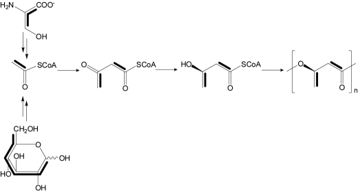
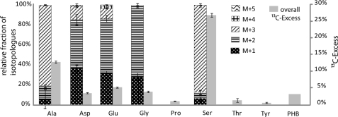
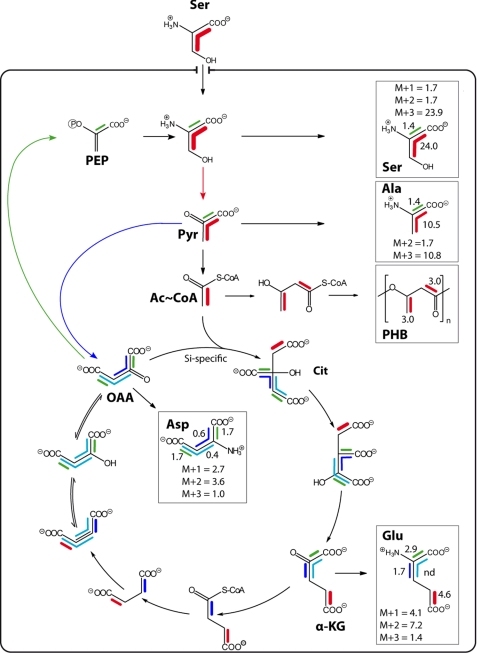
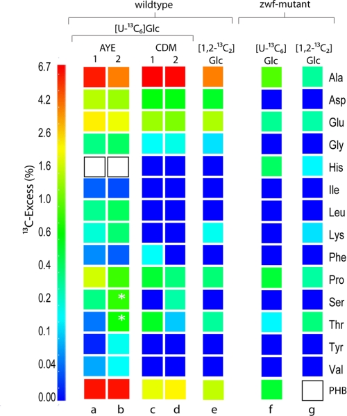
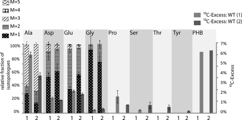
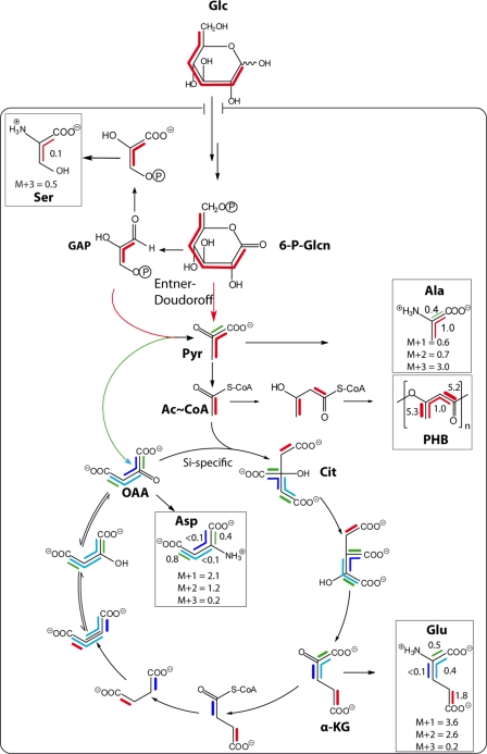
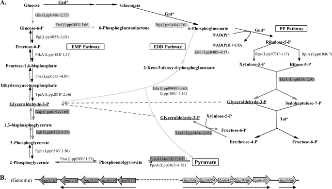
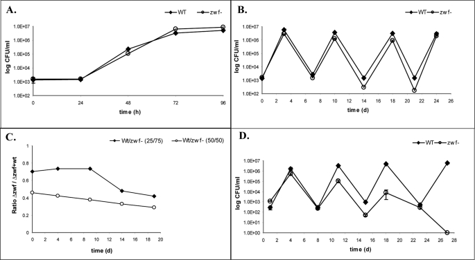
Legionella pneumophila (Lp) is commonly found in freshwater habitats but is also the causative agent of Legionnaires' disease when infecting humans. Although various virulence factors have been reported, little is known about the nutrition and the metabolism of the bacterium. Here, we report the application of isotopologue profiling for analyzing the metabolism of L. pneumophila. Cultures of Lp were supplied with [U-(13)C(3)]serine, [U-(13)C(6)]glucose, or [1,2-(13)C(2)]glucose. After growth, (13)C enrichments and isotopologue patterns of protein-derived amino acids and poly-3-hydroxybutyrate were determined by mass spectrometry and/or NMR spectroscopy. The labeling patterns detected in the experiment with [U-(13)C(3)]serine showed major carbon flux from serine to pyruvate and from pyruvate to acetyl-CoA, which serves as a precursor of poly-3-hydroxybutyrate or as a substrate of a complete citrate cycle with Si specificity of the citrate synthase. Minor carbon flux was observed between pyruvate and oxaloacetate/malate by carboxylation and decarboxylation, respectively. The apparent lack of label in Val, Ile, Leu, Pro, Phe, Met, Arg, and Tyr confirmed that L. pneumophila is auxotrophic for these amino acids. Experiments with [(13)C]glucose showed that the carbohydrate is also used as a substrate to feed the central metabolism. The specific labeling patterns due to [1,2-(13)C(2)]glucose identified the Entner-Doudoroff pathway as the predominant route for glucose utilization. In line with these observations, a mutant lacking glucose-6-phosphate dehydrogenase (Delta zwf) did not incorporate label from glucose at significant levels and was slowly outcompeted by the wild type strain in successive rounds of infection in Acanthamoeba castellanii, indicating the importance of this enzyme and of carbohydrate usage in general for the life cycle of Lp.


Figures

FIGURE 1.
FIGURE 1.
Formation of PHB in experiments with [U-13C3]Ser and [U-13C6]glucose. Multiple 13C labeling is indicated by bars connecting 13C-labeled atoms. The patterns of acetoacetyl-CoA and downstream products are overlays of isotopologues comprising two 13C-labeled atoms in a single molecule.
FIGURE 2.
FIGURE 2.
13C excess (gray columns; in mol % (right scale)) and isotopologue composition (patterned columns) of amino acids from the experiment with [U-13C3]Ser. The values represent means from three technical replicates, and the error bars indicate S.D. The patterned boxes indicate the relative contribution (% (left scale)) of 13C-isotopologues (M+1 to M+5 in different patterns) in the overall enrichments.
FIGURE 3.
FIGURE 3.
Metabolic model for Ser utilization by Lp Paris grown in culture. The scheme shows labeling patterns due to the incorporation of exogenous [U-13C3]Ser. Labeling patterns were detected in protein-derived amino acids and PHB (shown in boxes). Multiple 13C-labeled isotopologues determined by NMR spectroscopy are indicated as bars connecting 13C-labeled atoms in a given molecule. The numbers indicate the respective molar abundances. The molar abundances of isotopomer groups comprising one, two, or three 13C-labeled atoms, as determined by mass spectrometry (M+1, M+2, and M+3, respectively), are also listed in the boxes. PEP, phosphoenolpyruvate; OAA, oxaloacetate; α-KG, α-ketoglutarate.
FIGURE 4.
FIGURE 4.
Overall 13C excess (mol %) of labeled isotopologues in amino acids derived from protein after feeding of L. pneumophila Paris wild type and Δzwf mutant with 11 mm [U-13C6]glucose and 11 mm [1,2-13C2]glucose in different media. The color map indicates 13C excess in quasi-logarithmic form to show even relatively small 13C excess values. Each sample (different labeling experiments) was measured three times; the color for each amino acid correlates with the mean value of the three measurements. Asterisks indicate S.D. > 35%. PHB could not be measured in the experiment with [1,2-13C2]glucose and the Δzwf mutant.
FIGURE 5.
FIGURE 5.
13C excess (gray columns; in mol % (right scale)) and isotopologue composition (patterned columns) of amino acids from experiments 1 and 2 with L. pneumophila wild type cultivated in AYE medium containing 11 mm [U-13C6]glucose. The values represent means from three measurements, and error bars indicate S.D. The patterned boxes indicate the relative contribution (% (left scale)) of 13C-isotopologues (M+1 to M+5) in the overall enrichments.
FIGURE 6.
FIGURE 6.
Metabolic model for glucose utilization in Lp Paris grown in AYE medium supplemented with [U-13C6]glucose. Labeling patterns were detected in protein-derived amino acids and PHB (shown in boxes). Multiple 13C-labeled isotopologues determined by NMR spectroscopy are indicated as bars connecting 13C-labeled atoms in a given molecule. The numbers indicate the respective molar abundances. The molar abundances of isotopomer groups comprising one, two, or three 13C-labeled atoms as determined by mass spectrometry (M+1, M+2, and M+3, respectively) are also listed for comparison. GAP, glyceraldehyde-3-phosphate dehydrogenase; 6-P-Glcn, 6-phosphogluconate; Pyr, pyruvate; OAA, oxaloacetate; α-KG, α-ketoglutarate.
FIGURE 7.
FIGURE 7.
A, proposed EMP, ED, and PP pathways. The names of putative enzymes are shown; beneath them are the encoding open reading frames of Lp Paris (lpp), and the FC value of microarray analysis in vitro (28) are given in parentheses. FC, fold-change values (exponential phase versus stationary phase). Enzymes marked with an asterisk have no annotated homologues in the Lp genome. Genes determined to be co-transcribed are highlighted in gray and dark gray, respectively. Glk, glucokinase; Pgi, phosphoglucose isomerase; Pfk, phosphofructokinase; Fba, fructose-bisphosphate aldolase; TpiA, triose-phosphate isomerase; Gap, glyceraldehyde-3-phosphate dehydrogenase; Pgk, phosphoglycerate kinase; Pgm, phosphoglycerate mutase; Eno, enolase; PykA, pyruvate kinase; Zwf, glucose-6-phosphate dehydrogenase; Pgl, phosphogluconolactonase; Edd, phosphogluconate dehydratase; Eda, 2-keto-3-deoxy-phosphogluconate aldolase; Gnd, 6-phosphogluconate dehydrogenase; Rpe, ribulose phosphate-3-epimerase; RpiA, ribose-5-phosphate isomerase; TktA, transketolase; Tal, transaldolase; Gcd, glucose dehydrogenase; Gnt, gluconate transporter (modified from Ref. 16). B, schematic overview of the genes lpp043–lpp0488 (right) and lpp0150–lpp0154 (left). mRNA transcripts were determined via RT-PCR. lpp0483, zwf, lpp0484, pgl; lpp0485, edd; lpp0486, glk; lpp0487, eda; lpp0488 (putative sugar transport protein); lpp0150, sdhB (substrate of the Dot/Icm system); lpp0151, pykA; lpp0152, pgk; lpp0153, gap; lpp0154, tktA.
FIGURE 8.
FIGURE 8.
Analysis of L. pneumophila Paris (WT) and Δzwf mutant strain (zwf−) in co-cultures with A. castellanii. Bacteria were used to infect monolayers of A. castellanii at a multiplicity of infection of 0.01 with (A) Lp Paris or the Δzwf mutant strain for 96 h (A); Lp Paris or Δzwf mutant strain for 3 days, resuspended and incubated for a further 3 days, diluted to ∼103 bacteria/ml, and used to infect fresh amoebae (B). Four rounds of infection were performed. C, L. pneumophila Δzwf and WT strain survival over a 20-day period after co-infection of A. castellanii cells at two different ratios (circles, 50:50; diamonds, 75:25). D, infection was done as described in B, but A. castellanii cells were infected with both strains (WT and Δzwf) at the same time (in competition). At various time points postinoculation, bacteria were quantitated by plating aliquots on BCYE agar (see “Experimental Procedures”). Results are means ± S.D. of duplicate samples and are representative of at least three independent experiments.

Similar articles

Cited by

References

    1. Garduño R. A., Garduño E., Hiltz M., Hoffman P. S. (2002) Infect. Immun. 70, 6273–6283 - PMC - PubMed
    1. Hammer B. K., Tateda E. S., Swanson M. S. (2002) Mol. Microbiol. 44, 107–118 - PubMed
    1. Molofsky A. B., Swanson M. S. (2003) Mol. Microbiol. 50, 445–461 - PubMed
    1. Molofsky A. B., Swanson M. S. (2004) Mol. Microbiol. 53, 29–40 - PubMed
    1. Swanson M. S., Hammer B. K. (2000) Annu. Rev. Microbiol. 54, 567–613 - PubMed

Publication types

MeSH terms

LinkOut - more resources