. 2010 Jul 9;285(28):21292-302.
doi: 10.1074/jbc.M110.129874. Epub 2010 May 4.

R-spondin-1 is a novel beta-cell growth factor and insulin secretagogue

Affiliations

R-spondin-1 is a novel beta-cell growth factor and insulin secretagogue

Victor S C Wong et al. J Biol Chem. .

Abstract

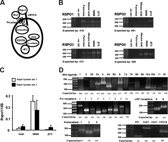
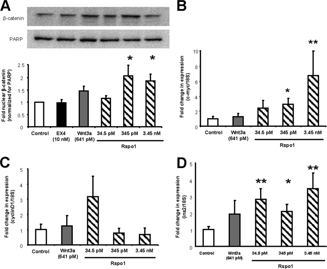
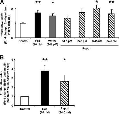
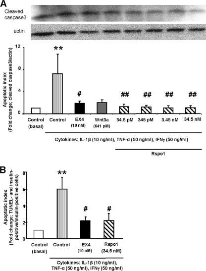
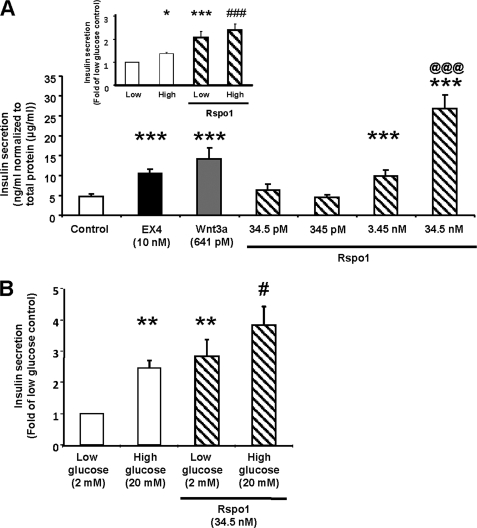
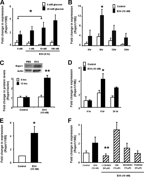
R-spondin-1 (Rspo1) is an intestinal growth factor known to exert its effects through activation of the canonical Wnt (cWnt) signaling pathway and subsequent expression of cWnt target genes. We have detected Rspo1 mRNA in murine islets and the murine MIN6 and betaTC beta-cell lines, and Rspo1 protein in MIN6 beta-cells. Rspo1 activated cWnt signaling in MIN6 beta-cells by increasing nuclear beta-catenin and c-myc, a cWnt target gene. Rspo1 also induced insulin mRNA expression in MIN6 cells. Analysis of MIN6 and mouse beta-cell proliferation by [(3)H]thymidine and BrdU incorporation, respectively, revealed that Rspo1 stimulated cell growth. Incubation of MIN6 and mouse beta-cells with cytokines (IL1beta/TNFalpha/interferon-gamma) significantly increased cellular apoptosis; this increase was abolished by pretreatment with Rspo1. Rspo1 also stimulated insulin secretion in a glucose-independent fashion. We further demonstrated that the glucagon-like peptide-1 receptor agonist, exendin4 (EX4), stimulated Rspo1 mRNA transcript levels in MIN6 cells in a glucose-, time-, dose-, and PI3-kinase-dependent fashion. This effect was not limited to this beta-cell line, as similar time-dependent increases in Rspo1 were also observed in the betaTC beta-cell line and mouse islets in response to EX4 treatment. Together, these studies demonstrate that Rspo1 is a novel beta-cell growth factor and insulin secretagogue that is regulated by EX4. These findings suggest that Rspo1 and the cWnt signaling pathway may serve as a novel target to enhance beta-cell growth and function in patients with type 2 diabetes.


Figures

FIGURE 1.
FIGURE 1.
Rspo1 and cWnt signaling molecules are expressed in murine β-cells. A, simplified schematic of cWnt and Rspo1 signaling showing cWnt ligand and Rspo1 binding to the Frz receptor and LRP5/6 co-receptor, as well as the intracellular protein Dishevelled and the β-catenin degradation complex consisting of APC, Axin, and GSK3β. B, RT-PCR analysis of Rspo1–4 mRNA in murine pancreas, islets, and MIN6 and βTC β-cell lines. A 100–1000-bp ladder was used. No RNA was used in the negative (-ve) control. C, relative qRT-PCR quantification of Rspo1 mRNA transcripts in murine islets, and MIN6 and βTC β-cells. Primer set 1, exons 2–3 and primer set 2, exons 3–4. Relative expression levels of Rspo1 were normalized to 18 S rRNA expression. (n = 5–30). D, RT-PCR for mRNA transcripts of various cWnt signaling molecules in MIN6 β-cells. A 100–1000-bp ladder was used.
FIGURE 2.
FIGURE 2.
Rspo1 activates cWnt signaling and increases insulin mRNA levels in MIN6 β-cells. A, ratio of nuclear β-catenin to nuclear PARP in MIN6 cells treated with EX4, Wnt3a, and increasing doses of Rspo1 for 30 min. A representative blot is shown. All values are expressed as fold-relative to the control (medium alone, n = 4–6). B–D, relative expression analysis of c-myc (B), cyclin D1 (C), and insulin (D) mRNA levels by qRT-PCR in MIN6 β-cells treated with medium alone (control), Wnt3a, or increasing doses of Rspo1 for 12 h. Data were normalized to the housekeeping gene 18 S rRNA (n = 9–11) and are displayed relative to vehicle-treated controls. *, p < 0.05 and **, p < 0.01.
FIGURE 3.
FIGURE 3.
Rspo1 stimulates β-cell proliferation. A, MIN6 β-cells were treated with either medium alone (control), EX4, Wnt3a or increasing doses of Rspo1 overnight, and their proliferation index was determined by [3H]thymidine incorporation assay (n = 14–33). B, dispersed murine islet cells were treated with medium alone (control), EX4 or Rspo1for 48 h, and BrdU was added for the last 24 h. Cells were then fixed and co-stained for insulin and BrdU. Proliferative index was determined as the number of BrdU- and insulin-positive cells over total insulin-positive cells, and data are presented as fold of control (n = 4). *, p < 0.05; **, p < 0.01.
FIGURE 4.
FIGURE 4.
Rspo1 inhibits cytokine-induced β-cell apoptosis. A and B, effects of Rspo1 on activated, cleaved caspase-3 in MIN6 β-cells (A) or TUNEL in dispersed murine β-cells (B). Cells were incubated in serum-free medium overnight, pretreated with medium alone (control), EX4, Wnt3a, or the specified doses of Rspo1 for 18 h, and then incubated without (basal) or with a combined cytokine mixture for a further 18 h. MIN6 β-cells were analyzed by immunoblotting for cleaved caspase-3 and pan-actin (n = 4–8). A representative blot is shown. Dispersed islet cells were fixed, and then co-stained for insulin and TUNEL (n = 6). Apoptotic index was expressed as fold change relative to the basal control group. *, p < 0.05 and **, p < 0.01 when compared with control (basal); #, p < 0.05 and ##, p < 0.01 when compared with control + cytokines.
FIGURE 5.
FIGURE 5.
Rspo1 stimulates insulin secretion in MIN6 β-cells and isolated mouse islets. A, insulin secretory response to Rspo1 in MIN6 β-cells (n = 5 - 12) was tested by static incubation of medium containing medium alone (control), EX4, Wnt3a, or indicated doses of Rspo1 for 2 h with high glucose. Insulin in the medium was measured by radioimmunoassay, and the results were normalized to total protein content. (inset: MIN6 β-cells were treated with or without Rspo1 (34.5 nm) under low (2 mm) or high glucose (25 mm) conditions (n = 6–12). Data were normalized to total protein content and expressed as fold of low glucose alone). B, insulin secretion in isolated mouse islets was determined after 2 h incubation with low or high glucose and with or without Rspo1. The results were normalized to total protein content and expressed as fold of low glucose alone. *, p < 0.05; **, p < 0.01; ***, p < 0.001 compared with control values, @@@, p < 0.001 for 34.5 nm compared with 3.45 nm Rspo1, and #, p < 0.05 and ###, p < 0.001 for high glucose compared with high glucose in presence of 34.5 nm Rspo1.
FIGURE 6.
FIGURE 6.
Rspo1 is regulated by EX4 in the β-cell. A, MIN6 β-cells were treated with EX4 at indicated concentrations under low or high glucose conditions for 8 h. mRNA levels of Rspo1 were examined by relative qRT-PCR using 18 S as the internal control and then normalized to control (5 mm glucose without EX4 at the 0 h time point). B, MIN6 β-cells were incubated in high glucose conditions with medium alone (control) or EX4 for the indicated times. C, protein levels of Rspo1 and actin were determined by immunoblot of MIN6 β-cells treated with medium alone (control) or EX4 for 8 or 12 h. Optical densities of Rspo1 were normalized to that of pan-actin and were further normalized to their appropriate controls. A representative blot is shown for the 12-h time point. D, qRT-PCR for Rspo1 mRNA expression in βTC β-cells after incubation with medium alone (control) or EX4 for the indicated times. Relative expression values were normalized 18 S rRNA and then to the 4 h control group. E, qRT-PCR for Rspo1 mRNA levels in mouse islets after incubation with medium alone (control) or EX4 for 4 h. Relative expression values were normalized to the control group. F, MIN6 β-cells were treated with or without EX4 and with various inhibitors, as indicated, for 8 h. Relative expression values for Rspo1 were normalized to 18 S rRNA and then to the control treatment. *, p < 0.05; **, p < 0.01.

Similar articles

Cited by

References

    1. Butler A. E., Janson J., Bonner-Weir S., Ritzel R., Rizza R. A., Butler P. C. (2003) Diabetes 52, 102–110 - PubMed
    1. Welters H. J., Kulkarni R. N. (2008) Trends Endocrinol. Metab. 19, 349–355 - PubMed
    1. Nusse R. (2005) Cell Res. 15, 28–32 - PubMed
    1. Fujino T., Asaba H., Kang M. J., Ikeda Y., Sone H., Takada S., Kim D. H., Ioka R. X., Ono M., Tomoyori H., Okubo M., Murase T., Kamataki A., Yamamoto J., Magoori K., Takahashi S., Miyamoto Y., Oishi H., Nose M., Okazaki M., Usui S., Imaizumi K., Yanagisawa M., Sakai J., Yamamoto T. T. (2003) Proc. Natl. Acad. Sci. U.S.A. 100, 229–234 - PMC - PubMed
    1. Schinner S., Ulgen F., Papewalis C., Schott M., Woelk A., Vidal-Puig A., Scherbaum W. A. (2008) Diabetologia 51, 147–154 - PubMed

Publication types