. 2010 Jul 9;285(28):21526-36.
doi: 10.1074/jbc.M110.129999. Epub 2010 May 4.

Knock-in mutation reveals an essential role for focal adhesion kinase activity in blood vessel morphogenesis and cell motility-polarity but not cell proliferation

Affiliations

Knock-in mutation reveals an essential role for focal adhesion kinase activity in blood vessel morphogenesis and cell motility-polarity but not cell proliferation

Ssang-Taek Lim et al. J Biol Chem. .

Abstract

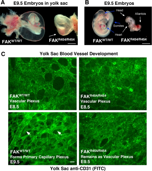
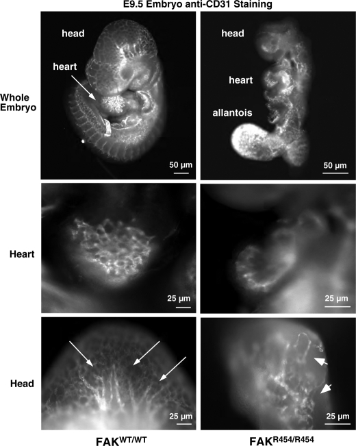
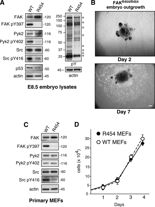
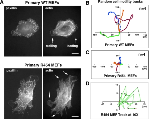
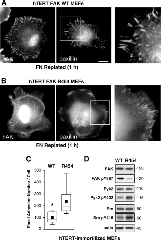
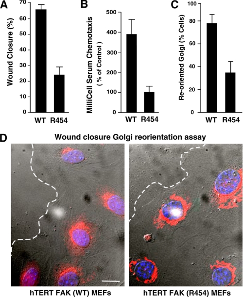
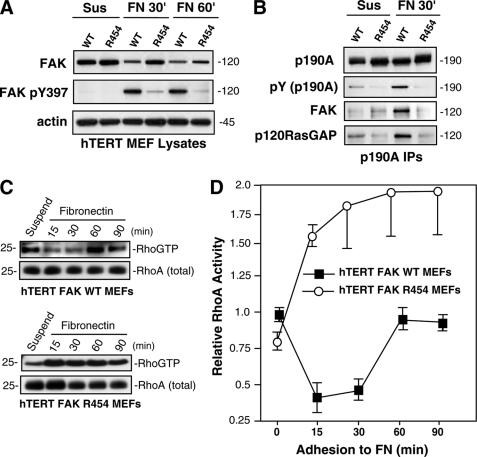
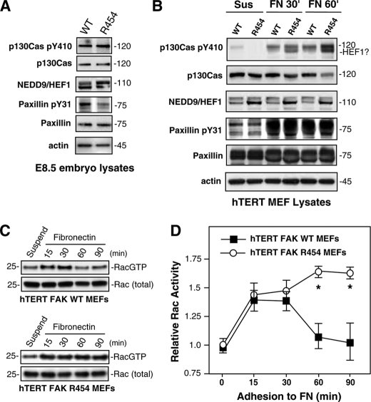
Focal adhesion kinase (FAK) associates with both integrins and growth factor receptors in the control of cell motility and survival. Loss of FAK during mouse development results in lethality at embryonic day 8.5 (E8.5) and a block in cell proliferation. Because FAK serves as both a scaffold and signaling protein, gene knock-outs do not provide mechanistic insights in distinguishing between these modes of FAK function. To determine the role of FAK activity during development, a knock-in point mutation (lysine 454 to arginine (R454)) within the catalytic domain was introduced by homologous recombination. Homozygous FAK(R454/R454) mutation was lethal at E9.5 with defects in blood vessel formation as determined by lack of yolk sac primary capillary plexus formation and disorganized endothelial cell patterning in FAK(R454/R454) embryos. In contrast to the inability of embryonic FAK(-/-) cells to proliferate ex vivo, primary FAK(R454/R454) mouse embryo fibroblasts (MEFs) were established from E8.5 embryos. R454 MEFs exhibited no difference in cell growth compared with normal MEFs, and R454 FAK localized to focal adhesions but was not phosphorylated at Tyr-397. In E8.5 embryos and primary MEFs, FAK R454 mutation resulted in decreased c-Src Tyr-416 phosphorylation. R454 MEFs exhibited enhanced focal adhesion formation, decreased migration, and defects in cell polarity. Within immortalized MEFs, FAK activity was required for fibronectin-stimulated FAK-p190RhoGAP association and p190RhoGAP tyrosine phosphorylation linked to decreased RhoA GTPase activity, focal adhesion turnover, and directional motility. Our results establish that intrinsic FAK activity is essential for developmental processes controlling blood vessel formation and cell motility-polarity but not cell proliferation. This work supports the use of FAK inhibitors to disrupt neovascularization.


Figures

FIGURE 1.
FIGURE 1.
FAKR454/R454 kinase-inactive knock-in mutation is embryonic lethal with vascular defects. A, bright field images of excised yolk sacs with embryos reveals a lack of blood-filled vessels on FAKR454/R454 compared with FAKWT/WT embryos at E9.5. The red color within the FAKR454/R454 yolk sac (arrow) is associated with internal embryo hemorrhage. Bar, 100 μm. B, at E9.5, FAKWT/WT embryos have undergone turning and have a defined head, beating heart, and multiple segmental somites (arrows). FAKR454/R454 embryos at E9.5 have undergone turning, are small, and possess head and heart structures that are malformed (arrows). No somites or beating hearts were detectable in FAKR454/R454 embryos. The allantois is enlarged, does not undergo chorion fusion, and is frequently a site of hemorrhage within FAKR454/R454 embryos. Bar, 100 μm. C, whole mount anti-CD31 staining of yolk sacs at E8.5 reveals equivalent vascular plexus formation in FAKR454/R454 and FAKWT/WT embryos. At E9.5, ECs within yolk sacs of FAKWT/WT embryos form a primary capillary plexus characterized by tubules and branched structures (arrowheads). The primary capillary plexus is not formed in FAKR454/R454 yolk sacs at E9.5 because this remains a vascular plexus structure. Bar, 20 μm.
FIGURE 2.
FIGURE 2.
Defective EC patterning within FAKR454/R454 embryos. Whole embryos at E9.5 were stained with anti-CD31 to visualize ECs. Comparisons are between FAKWT/WT and FAKR454/R454 littermates. Defined EC branchlike and tubule structures (arrows) in the head and heart of FAKWT/WT embryos are shown. FAKR454/R454 embryos exhibit intense EC staining in the unfused allantois. Clusters (arrowheads) of ECs are present in FAKR454/R454 head and heart structures, but these were not organized within a defined network.
FIGURE 3.
FIGURE 3.
R454 FAK expression and FAK phosphorylation target identification within E8.5 embryo lysates and establishment of primary FAKR454/R454 MEFs. A and C, protein lysates from FAKWT/WT (WT) and FAKR454/R454 (R454) embryos harvested at E8.5 or lysates from primary MEFs were analyzed by immunoblotting for total FAK, Tyr(P)-397 (pY397) FAK, total Pyk2, Pyk2 Tyr(P)-402 (pY402), total c-Src, and Src Tyr(P)-416 (pY416). Anti-phosphotyrosine (pY) immunoblotting revealed decreased target protein tyrosine phosphorylation (asterisks) in lysates from E8.5 R454 embryos. Anti-actin immunoblotting was used as a loading control. Decreased c-Src activation (as detected by Src Tyr(P)-416 blotting) was detected in R454 embryos and MEFs. A, p53 tumor suppressor expression was lower in R454 E8.5 embryos as determined by immunoblotting. B, phase-contrast images of E8.5 FAKR454/R454 embryo outgrowth in culture after 2 and 7 days. Shown is Matrigel-embedded embryo (dark center) and surrounding cell outgrowth. Bar, 100 μm. D, primary WT and R454 MEFs proliferate equally in culture. Results are the mean cell number (n = 3 independent points ± S.D. (error bars)).
FIGURE 4.
FIGURE 4.
Primary R454 MEFs exhibit increased FA formation and lack directional persistence of cell movement. A, WT and R454 MEFs were plated onto FN-coated glass slides for 1 h and co-stained for paxillin and actin. WT MEFs formed defined leading and trailing edge cell protrusions, whereas R454 MEFs formed protrusions randomly around the cell periphery (arrows). Increased numbers of paxillin-stained FAs formed in R454 MEFs. Bar, 10 μm. B, analysis of WT MEF migration paths by time lapse imaging of MEFs plated on FN in the presence of serum. Cell tracks were determined by cell nuclei position, and migration origin was superimposed at 0. The scale is in μm, n = 4 cells. C, analysis of R454 MEF migration paths as in B. The scale is μm, n = 4 cells. D, ×10 magnified migration track from C shows the lack of directionality of R454 MEF movement.
FIGURE 5.
FIGURE 5.
Immortalized FAK R454 MEFs exhibit increased FA formation. Primary WT and R454 MEFs were immortalized by retroviral hTERT expression, and pooled populations of cells were characterized. A and B, MEFs were plated on FN for 1 h in the absence of serum and stained with antibodies to FAK and paxillin. Bar, 10 μm. Inset (right), enlargement of paxillin-stained FAs. C, increased FA formation in R454 MEFs. Box-and-whisker plots of paxillin-positive stained points were enumerated within WT and R454 MEFs plated on FN for 1 h (n = 15 cells/point). Box-and-whisker diagrams show the distribution of the data: mean (square), 25th percentile (bottom line), median (middle line), 75th percentile (top line), and 5th or 95th percentiles (whiskers). Significance was determined using a two-tailed Student's t test (*, p < 0.0001). D, lysates from WT and R454 MEFs were by analyzed by total FAK, Tyr(P)-397 (pY397) FAK, total Pyk2, Pyk2 Tyr(P)-402 (pY402), total c-Src, Src Tyr(P)-416 (pY416), and actin immunoblotting. R454 FAK is expressed but only weakly phosphorylated at Tyr-397. Increased levels of Pyk2 and c-Src activation were detected in hTERT-immortalized R454 compared with WT MEFs.
FIGURE 6.
FIGURE 6.
Immortalized FAK R454 MEFs exhibit motility and polarity defects. A, wound closure motility of WT and R454 MEFs was calculated by measuring the distance change between 0 and 16 h in time lapse experiments. Values are means ± S.D. from 10 independent analyses. B, chamber motility was measured over 4 h with WT and R454 MEFs with FN and serum. Values are means ± S.D. from three experiments. C, FAK activity is required for Golgi reorientation and cell polarization. 100 cells were analyzed, and values represent percentage of cells ± S.D. D, representative images from Golgi reorientation assay. WT and R454 MEFs were grown to confluence, wounded with a pipette tip, and allowed to migrate in the presence of serum for 4 h. Cells were fixed, imaged in phase, and stained for Golgi (β-Cop, red) and nuclear (Hoechst, blue) markers. Position of the leading lamella (dashed line) is indicated. Bar, 10 μm.
FIGURE 7.
FIGURE 7.
FAK promotes p190RhoGAP phosphorylation after FN stimulation of MEFs; connection to RhoA regulation. A, R454 FAK is not activated upon MEF adhesion to FN. Lysates from suspended and FN-replated (30 and 60 min) hTERT WT and R454 MEFs were analyzed by total FAK, Tyr(P)-397 (pY397) FAK, and actin blotting. B, p190A forms a complex with FAK and p120RasGAP upon WT but not R454 MEF adhesion to FN. p190A immunoprecipitations were made from lysates of hTERT WT or R454 MEFs held in suspension or FN-replated (30 min) and sequentially analyzed by anti-Tyr(P) (pY) and -p190A blotting. The 120 kDa region of the p190A immunoprecipitations (IPs) was sequentially analyzed by anti-FAK and -120RasGAP immunoblotting. C, transient decrease in RhoA GTP binding upon WT but not R454 MEF adhesion to FN. GTP-bound RhoA was determined by GST-rhotekin Rho-binding domain affinity pull-down assays from lysates of suspended and FN-replated cells, followed by blotting for total RhoA levels. D, elevated RhoA GTP binding was observed in R454 FAK MEFs upon FN adhesion. Quantitation of RhoA-GTP binding from two independent experiments ± S.D. Values are normalized to total RhoA levels are relative to RhoA-GTP levels in suspended WT FAK MEFs.
FIGURE 8.
FIGURE 8.
Elevated Rac GTP levels in FAK R454 MEFs upon FN replating are associated with increased HEF1 expression and p130Cas tyrosine phosphorylation. Protein lysates from FAKWT/WT (WT) and FAKR454/R454 (R454) embryos harvested at E8.5 (A) or protein lysates from hTERT-immortalized WT or R454 MEFs held in suspension for 30 min (Sus) or replated onto fibronectin (FN) for 30 or 60 min in the absence of serum (B) were evaluated by immunoblotting for total p130Cas, Tyr(P)-410 (pY410) p130Cas, NEDD9/HEF1, total paxillin, Tyr(P)-31 (pY31) paxillin, and actin. C, elevated GTP-bound Rac1 in R454 MEFs upon FN adhesion. GTP-bound Rac1 was determined by affinity pull-down assays (GST-PAK residues 67–150) from lysates of suspended and fibronectin-replated WT and R454 FAK hTERT-immortalized MEFs followed by blotting for total Rac1 levels. D, quantitation of Rac1-GTP binding and values normalized to total Rac1 levels and plotting relative to Rac1-GTP levels in suspended WT FAK MEFs. Data are from two independent experiments ± S.D. Significance was determined by two-way analysis of variance (*, p < 0.01).

Similar articles

Cited by

References

    1. Serini G., Valdembri D., Bussolino F. (2006) Exp. Cell Res. 312, 651–658 - PubMed
    1. Jain R. K. (2003) Nat. Med. 9, 685–693 - PubMed
    1. Risau W. (1997) Nature 386, 671–674 - PubMed
    1. Hynes R. O. (2007) J. Thromb. Haemost. 5, Suppl. 1, 32–40 - PubMed
    1. Giancotti F. G., Ruoslahti E. (1999) Science 285, 1028–1032 - PubMed

Publication types

MeSH terms

Substances