. 2010 Jul 2;285(27):20595-606.
doi: 10.1074/jbc.M109.092411. Epub 2010 May 4.

Protein-arginine methyltransferase 1 suppresses megakaryocytic differentiation via modulation of the p38 MAPK pathway in K562 cells

Affiliations

Protein-arginine methyltransferase 1 suppresses megakaryocytic differentiation via modulation of the p38 MAPK pathway in K562 cells

Yuan-I Chang et al. J Biol Chem. .

Abstract

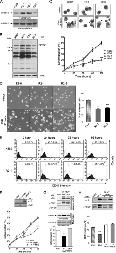
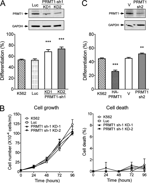
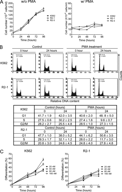
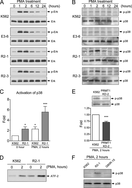
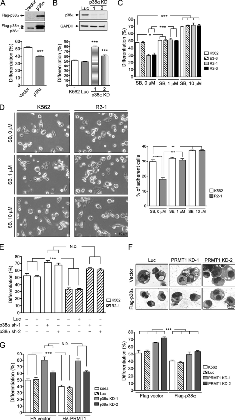
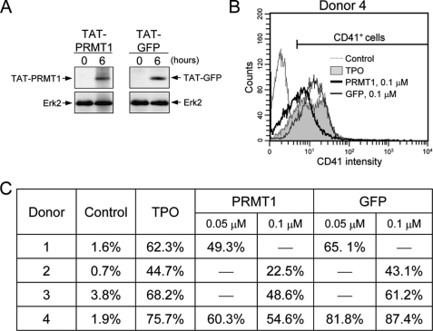
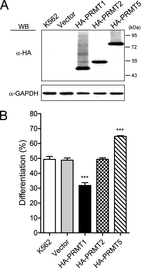
Protein-arginine methyltransferase 1 (PRMT1) plays pivotal roles in various cellular processes. However, its role in megakaryocytic differentiation has yet to be investigated. Human leukemia K562 cells have been used as a model to study hematopoietic differentiation. In this study, we report that ectopic expression of HA-PRMT1 in K562 cells suppressed phorbol 12-myristate 13-acetate (PMA)-induced megakaryocytic differentiation as demonstrated by changes in cytological characteristics, adhesive properties, and CD41 expression, whereas knockdown of PRMT1 by small interference RNA promoted differentiation. Impairment of the methyltransferase activity of PRMT1 diminished the suppressive effect. These results provide evidence for a novel role of PRMT1 in negative regulation of megakaryocytic differentiation. Activation of ERK MAPK has been shown to be essential for megakaryocytic differentiation, although the role of p38 MAPK is still poorly understood. We show that knockdown of p38alpha MAPK or treatment with the p38 inhibitor SB203580 significantly enhanced PMA-induced megakaryocytic differentiation. Further investigation revealed that PRMT1 promotes activation of p38 MAPK without inhibiting activation of ERK MAPK. In p38alpha knockdown cells, PRMT1 could no longer suppress differentiation. In contrast, enforced expression of p38alpha MAPK suppressed PMA-induced megakaryocytic differentiation of parental K562 as well as PRMT1-knockdown cells. We propose modulation of the p38 MAPK pathway by PRMT1 as a novel mechanism regulating megakaryocytic differentiation. This study thus provides a new perspective on the promotion of megakaryopoiesis.


Figures

FIGURE 1.
FIGURE 1.
Ectopic expression of HA-PRMT1 suppresses PMA-induced megakaryocytic differentiation of K562 cells. K562 cells were stably transfected with pPCDNA3HA2-PRMT1 plasmids (R2-1 and R2-3) or empty vectors (E3-6). The methyltransferase activity of PRMT1 in cell homogenates was assayed using the known substrate hnRNP K and detected by fluorography (A). Levels of asymmetric dimethylarginine, a product of PRMT1 activity, in cell lysates were detected by Western blot (WB) analysis using a specific antibody (ASYM24) (B). Various K562 cell clones were treated with PMA (40 nm). Megakaryocytic differentiation was detected by modified Wright-Giemsa staining for cytological changes (C), by phase contrast microscopy for examination of adherent cells with pseudopodia (D), or by flow cytometry for the expression of the specific surface marker CD41 (E). Cells with enlarged and lobed nuclei and microvesicles are marked by arrowheads (C, upper panel). The suspended and adherent cells were collected and quantified (D, right panel). Dashed lines in E are isotypic controls. Ectopic expression of HA-PRMT1, R2-1, and R2-3 significantly suppressed differentiation. Transient transfection of K562 cells with pPCDNA3HA2-PRMT1 led to similar effects (F). Inset shows expression of HA-PRMT1 in transfected cells. The enzymatically impaired mutant PRMT1G80R, when transiently expressed in K562 cells, did not suppress differentiation as the wild type (WT) enzyme (G, lower panel). Ectopic expression of mutant PRMT1 did not increase the methyltransferase activity in cell homogenates when assayed with a known PRMT1 substrate hnRNP K (G, middle panel). Expression levels of wild type and mutant HA-PRMT1 proteins were similar (G, upper panel). Stable clones of mutant PRMT1G80R also did not show suppressive effects on differentiation (H, lower panel). Expression of wild type and mutant HA-PRMT1 proteins were examined by Western blot (H, upper panel). All experiments were performed at least three times, and data are presented as means ± S.E.; *, p < 0.05; **, p < 0.01; ***, p < 0.005 as compared with K562 parental cells.
FIGURE 2.
FIGURE 2.
Reduced levels of endogenous PRMT1 enhance PMA-induced megakaryocytic differentiation of K562 cells. KD-1 and KD-2 cell clones were stably transfected with PRMT1 shRNA (sh-1). Western analysis using an anti-PRMT1 antibody showed reduced expression of PRMT1 proteins (A, upper panel). Megakaryocytic differentiation was analyzed 96 h after PMA treatment (A, lower panel). Luc, luciferase shRNA. The growth and viability of these cell clones were similar under regular culture conditions (B). Effects of PRMT1 knockdown were also assayed by transient transfection with a different shRNA (sh-2). PRMT1 protein levels were detected by Western blot analysis (C, upper panel) and megakaryocytic differentiation was detected by modified Wright-Giemsa staining (C, lower panel). Luciferase shRNA (Luc) and the pSUPER empty vector (V) were used as controls. All experiments were performed at least three times, and data are presented as means ± S.E.; **, p < 0.01; ***, p < 0.005 as compared with K562 parental cells.
FIGURE 3.
FIGURE 3.
Differential effects of PRMT1 on PMA-induced growth arrest and differentiation. Under regular culture conditions, the growth rate of HA-PRMT1-expressing cells, R2-1 and R2-3, were similar to parental cells (A, left panel). PMA treatment (40 nm) caused a cessation of growth (A, right panel) and cell cycle arrest at G1 phase (B) in all cell clones. To examine the dose response of these cells to PMA, K562 and R2-1 cells were treated with increasing concentrations of PMA as indicated, and megakaryocytic differentiation was analyzed by modified Wright-Giemsa staining (C). All experiments were performed at least three times and data are presented as means ± S.E. w/o, without; w/, with.
FIGURE 4.
FIGURE 4.
Modulation of the MAPK pathways by PRMT1. Parental, empty control (E3-6), and HA-PRMT1-expressing R2-1 and R2-3 cells were treated with PMA (40 nm), collected, and lysed in RIPA buffer. Activation of ERK (A) and p38 (B) was detected with antibodies against the specific phosphorylated forms. Activation of p38 2 h after PMA treatment was quantified and normalized to the total amount of p38 protein (C). The active form of p38 was immunoprecipitated, and its kinase activity was assayed by phosphorylation of its substrate ATF-2 (D). The kinase activity of p38 was significantly activated in PRMT1-overexpressing cells (C and D). Knockdown of endogenous PRMT1 decreased the activation of p38 (E). Abrogation of PRMT1 enzyme activity (G80R-15 mutant) could no longer promote activation of p38 (F). All experiments were performed at least three times, and data are presented as means ± S.E.; *, p < 0.05; ***, p < 0.005 as compared with K562 parental cells.
FIGURE 5.
FIGURE 5.
PRMT1-mediated suppression of megakaryocytic differentiation is dependent on activation of the p38 MAPK. K562 cells were transiently transfected with FLAG-p38α and empty vectors and analyzed for megakaryocytic differentiation 96 h after PMA treatment (A). Stable cell clones (p38α KD) transfected simultaneously with two p38α shRNAs (sh-1 and sh-2) were selected, and the protein levels were examined by Western blot analysis (B, upper panel). Luc, luciferase shRNA; GAPDH, glyceraldehyde-3-phosphate dehydrogenase. Reduced expression of p38α enhanced megakaryocytic differentiation (B, lower panel). Treatment with the p38 inhibitor SB203580 (SB) greatly enhanced megakaryocytic differentiation in both parental K562 and HA-PRMT1-expressing R2-1 and R2-3 cells when analyzed 96 h after PMA treatment (C). The specific inhibitor of p38 MAPK, SB203580, was added 30 min before stimulation with PMA. Adherent cells were examined by phase contrast microscopy 96 h after PMA treatment (D, left panel) and quantified (D, right panel). Transient knockdown of p38α with either p38α sh-1 or p38α sh-2 shRNAs enhanced megakaryocytic differentiation in both K562 and R2-1 cells (E). Ectopic expression of p38α by transient transfection suppressed megakaryocytic differentiation in both K562 and PRMT1-knockdown cells (PRMT1 KD-1 and KD-2) (F). Cytological changes were detected by modified Wright-Giemsa staining (F, upper panel) and quantified (F, lower panel). HA-PRMT1 was transiently expressed in stable p38α-knockdown clones (p38α KD-1 and p38α KD-2) (G). Ectopic expression of HA-PRMT1 could no longer suppress megakaryocytic differentiation in p38α-deficient cells. Cells stably transfected with the luciferase shRNA (Luc) were used as a negative control. All experiments were performed at least three times, and data are presented as means ± S.E.; *, p < 0.05; **, p < 0.01; ***, p < 0.005; N.D. means no difference.
FIGURE 6.
FIGURE 6.
Enforced expression of PRMT1 in human CD34+ hematopoietic cells suppresses TPO-induced megakaryocytic differentiation. Recombinant TAT-HA-PRMT1 and TAT-HA-GFP (control) proteins were added to CD34+ cells. Entrance of the TAT-fused proteins into cells was examined by Western blot using anti-HA antibodies (A). ERK2 was used as a loading control. To examine the effect on megakaryocytic differentiation, TAT-fused proteins were added into cells 6 h before TPO treatment and again at the time of TPO treatment. After 15 days, cells were analyzed for expression of CD41 by flow cytometry (B and C). A representative result from donor 4 is shown in B.
FIGURE 7.
FIGURE 7.
Effect of PRMT2 and PRMT5 on megakaryocytic differentiation. K562 cells were transiently transfected with pPCDNA3HA2 plasmids expressing either HA-tagged PRMT1, PRMT2, or PRMT5 proteins that were detected by Western blot (WB) using anti-HA antibodies (A). Glyceraldehyde-3-phosphate dehydrogenase (GAPDH) was used as a loading control. Megakaryocytic differentiation was analyzed by modified Wright-Giemsa staining 96 h after PMA treatment (B). All experiments were performed at least three times, and data are presented as means ± S.E.; ***, p < 0.005.

Similar articles

Cited by

References

    1. Kaushansky K. (2005) J. Clin. Invest. 115, 3339–3347 - PMC - PubMed
    1. Alitalo R. (1990) Leuk. Res. 14, 501–514 - PubMed
    1. Racke F. K., Lewandowska K., Goueli S., Goldfarb A. N. (1997) J. Biol. Chem. 272, 23366–23370 - PubMed
    1. Whalen A. M., Galasinski S. C., Shapiro P. S., Nahreini T. S., Ahn N. G. (1997) Mol. Cell. Biol. 17, 1947–1958 - PMC - PubMed
    1. Herrera R., Hubbell S., Decker S., Petruzzelli L. (1998) Exp. Cell Res. 238, 407–414 - PubMed

Publication types

MeSH terms

Substances