. 2010 Jun 2;29(11):1877-88.
doi: 10.1038/emboj.2010.80. Epub 2010 May 4.

Dynamic link of DNA demethylation, DNA strand breaks and repair in mouse zygotes

Affiliations

Dynamic link of DNA demethylation, DNA strand breaks and repair in mouse zygotes

Mark Wossidlo et al. EMBO J. .

Abstract

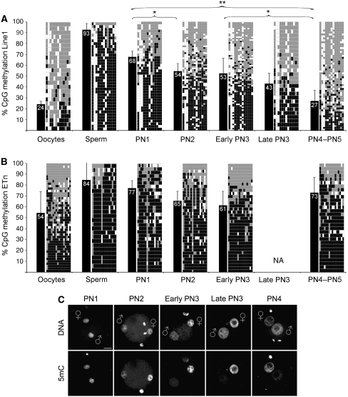
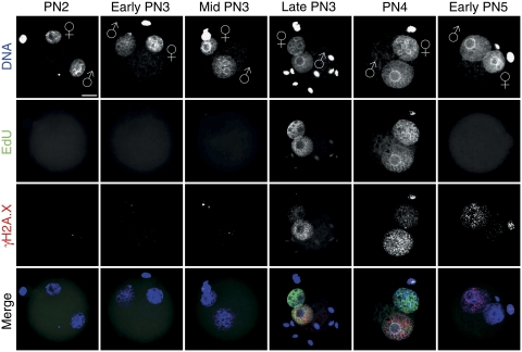
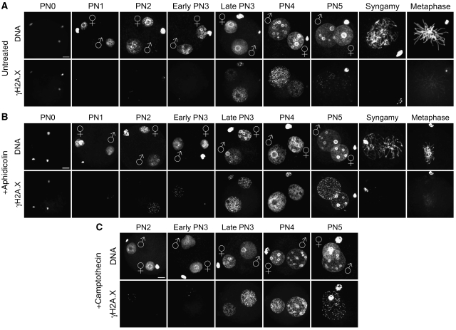
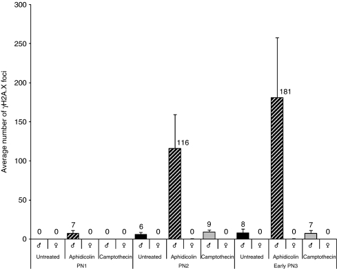
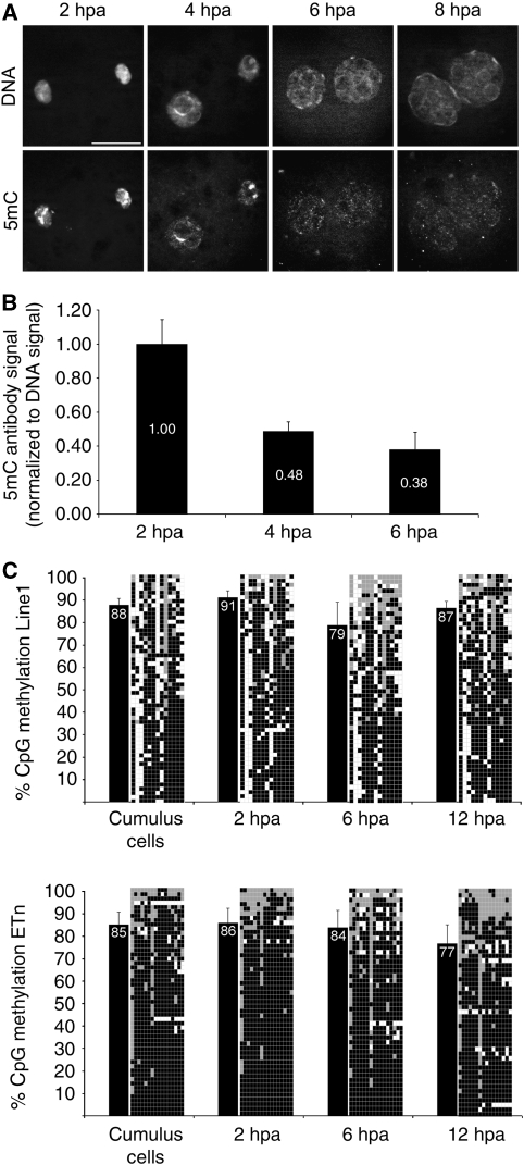
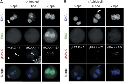
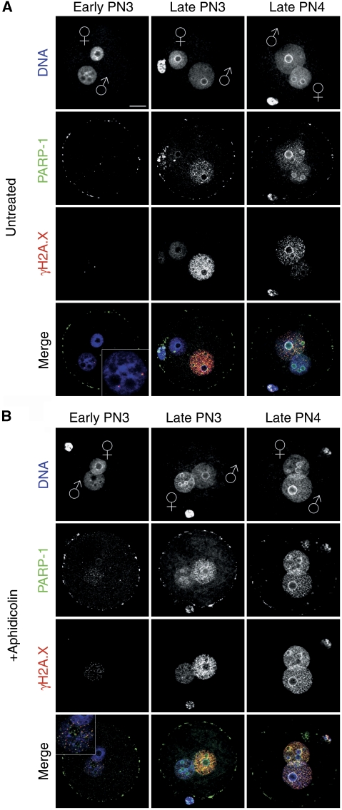
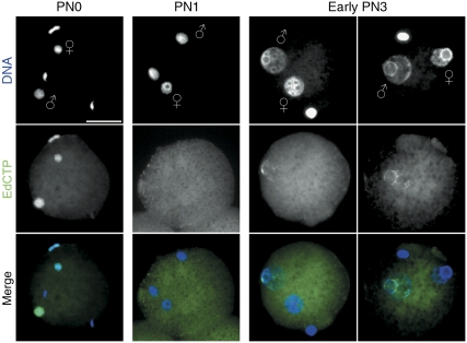
In mammalian zygotes, the 5-methyl-cytosine (5mC) content of paternal chromosomes is rapidly changed by a yet unknown but presumably active enzymatic mechanism. Here, we describe the developmental dynamics and parental asymmetries of DNA methylation in relation to the presence of DNA strand breaks, DNA repair markers and a precise timing of zygotic DNA replication. The analysis shows that distinct pre-replicative (active) and replicative (active and passive) phases of DNA demethylation can be observed. These phases of DNA demethylation are concomitant with the appearance of DNA strand breaks and DNA repair markers such as gammaH2A.X and PARP-1, respectively. The same correlations are found in cloned embryos obtained after somatic cell nuclear transfer. Together, the data suggest that (1) DNA-methylation reprogramming is more complex and extended as anticipated earlier and (2) the DNA demethylation, particularly the rapid loss of 5mC in paternal DNA, is likely to be linked to DNA repair mechanisms.


Conflict of interest statement

The authors declare that they have no conflict of interest.

Figures

Figure 1
Figure 1
Line1/ETn CpG methylation and loss of 5mC antibody signal during zygotic development. (A) Line1 CpG methylation of gametes and zygotes at distinct pronuclear stages reveals a stepwise demethylation process. Black bars indicate average methylation status. Diagrams specify analysed CpG positions where each row depicts an individual chromosomal Line1 pattern (black=methylated, grey=unmethylated, white=not analysable/mutated). Note the increasing mosaicism of methylated CpG positions over time. Asterisks indicate significant methylation changes between different pronuclear stages. (B) ETn CpG methylation of gametes and zygotes at distinct pronuclear stages. The methylation level also decreases from PN1 up to early PN3 likewise Line1. After completion of S-phase, the average methylation level increases again. (C) Representative IF analysis with a monoclonal Ab against 5mC during pronuclear stages (see also Santos et al, 2002). The major loss of Ab signal in the paternal pronucleus occurs between PN2 and PN3. Scale bar=20 μm.
Figure 2
Figure 2
Timing of DNA replication in mouse zygotes. Representative images of click-iT-labelled EdU incorporation during pronuclear stages (classified according to Adenot et al, 1997) (n>60, at least three independent IVFs per PN stage). Pulse labelling of IVF zygotes with EdU was performed for 30 min before fixation. EdU incorporation occurs synchronously in male and female pronuclei at late PN3–PN4. The lower two panels show the co-staining with anti-γH2A.X and the merge with EdU signals, which depict the replication-associated γH2A.X foci formation predominantly in paternal pronuclei. Note the few pre-replicative γH2A.X foci in the paternal pronucleus and the stronger staining at early PN5. Scale bar=20 μm.
Figure 3
Figure 3
Dynamics of γH2A.X signal during zygotic development. Representative images of indirect immunostainings using antibodies against γH2A.X in IVF zygotes at distinct pronuclear stages (PN0–PN5, syngamy and metaphase). (A) γH2A.X dynamics in untreated controls. (B) IVF zygotes incubated with aphidicolin for 2 h before fixation. (C) IVF zygotes incubated with camptothecin for 2 h prior the fixation. Scale bar=20 μm. A colour version of this figure is available at The EMBO Journal Online (Supplementary Figure S2).
Figure 4
Figure 4
Number of γH2A.X foci in paternal and maternal pronuclei during G1-phase. The graph shows the average number of γH2A.X foci (n>20 zygotes/analysis) in both parental pronuclei in untreated, aphidicolin-treated and camptothecin-treated IVF zygotes. In aphidicolin-treated zygotes at G1-phase, a significant increase of γH2A.X foci can be detected only in paternal pronuclei.
Figure 5
Figure 5
Loss of 5mC-antibody signal and Line1/ETn methylation in cloned embryos. (A) Indirect immunostainings with antibodies against 5mC in SCNT embryos at 2, 4, 6 and 8 hpa. Note the dynamic loss of antibody signal in both pronuclei at 6 hpa. (B) Quantification of 5mC antibody signal normalized to DNA signal (n=4 per stage). At 4 hpa, the 5mC antibody signal decreases to 48% compared with 2 hpa. (C) Line1 bisulphite sequencing of cloned one-cell embryos at distinct pronuclear stages reveals only a minor drop of DNA methylation levels at 6 hpa, which then slightly increases at 12 hpa. ETn methylation levels remain rather constant with the tendency to decrease after replication. Black bars indicate average methylation status. Diagrams specify analysed CpG positions where each row depicts an individual chromosomal Line1 pattern (black=methylated, grey=unmethylated, white=not analysable/mutated).
Figure 6
Figure 6
Dynamics of γH2A.X signal in cloned embryos. (A) SCNT embryos incubated for 1 hour with EdU before fixation at 5, 6 and 7 hpa. S-phase starts at 7 hpa coinciding with the appearance of increased γH2A.X signals. (B) Aphidicolin blocks EdU incorporation and enhances the γH2A.X signal in both G1- (up to 6 hpa) and S-phase (7 hpa). Scale bar=20 μm; arrows indicate single γH2A.X foci.
Figure 7
Figure 7
PARP-1 co-localization with γH2A.X. Representative indirect immunostainings using antibodies against PARP-1 and γH2A.X in pre-replicative and replicative stages. The picture shows computed z-stacks to reduce unspecific background staining of PARP-1 immunostainings on the surface of the zygotes, which is still visible as ‘ring' on the oolemma. (A) PARP-1 co-localization with γH2A.X at pre-replicative and replicative pronuclear stages. (B) Aphidicolin treatment enhances the PARP-1 signal co-localized with also enhanced γH2A.X foci. Scale bar=20 μm.
Figure 8
Figure 8
DNA single-strand breaks in pre-replicative zygotes. Modified nick translation assay using the 5-ethynyl-2′-deoxycytidine-triphosphate (EdCTP) for click-iT fluorescence labelling. PolI-mediated incorporation of EdCTP in mildly decondensed G1-phase zygotes. At PN0, both parental genomes show the presence of SSBs, whereas at PN1 stage, no nicks can be detected. In early PN3 zygotes, the signal exclusively appears in paternal pronuclei (two examples). Scale bar=20 μm.

References

    1. Adenot PG, Mercier Y, Renard JP, Thompson EM (1997) Differential H4 acetylation of paternal and maternal chromatin precedes DNA replication and differential transcriptional activity in pronuclei of 1-cell mouse embryos. Development 124: 4615–4625 - PubMed
    1. Aoki E, Schultz RM (1999) DNA replication in the 1-cell mouse embryo: stimulatory effect of histone acetylation. Zygote 7: 165–172 - PubMed
    1. Aoki F, Worrad DM, Schultz RM (1997) Regulation of transcriptional activity during the first and second cell cycles in the preimplantation mouse embryo. Dev Biol 181: 296–307 - PubMed
    1. Barreto G, Schafer A, Marhold J, Stach D, Swaminathan SK, Handa V, Doderlein G, Maltry N, Wu W, Lyko F, Niehrs C (2007) Gadd45a promotes epigenetic gene activation by repair-mediated DNA demethylation. Nature 445: 671–675 - PubMed
    1. Berger NA, Kurohara KK, Petzold SJ, Sikorski GW (1979) Aphidicolin inhibits eukaryotic DNA replication and repair—implications for involvement of DNA polymerase alpha in both processes. Biochem Biophys Res Commun 89: 218–225 - PubMed

Publication types

LinkOut - more resources