. 2010 Jul;18(7):1379-88.
doi: 10.1038/mt.2010.75. Epub 2010 May 4.

FimH can directly activate human and murine natural killer cells via TLR4

Affiliations

FimH can directly activate human and murine natural killer cells via TLR4

M Firoz Mian et al. Mol Ther. 2010 Jul.

Abstract

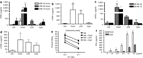
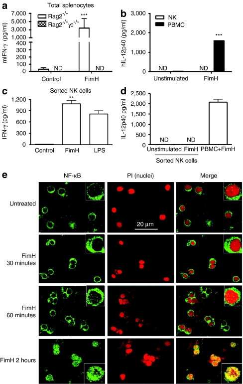
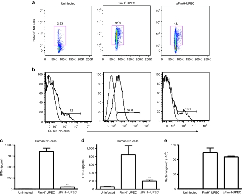
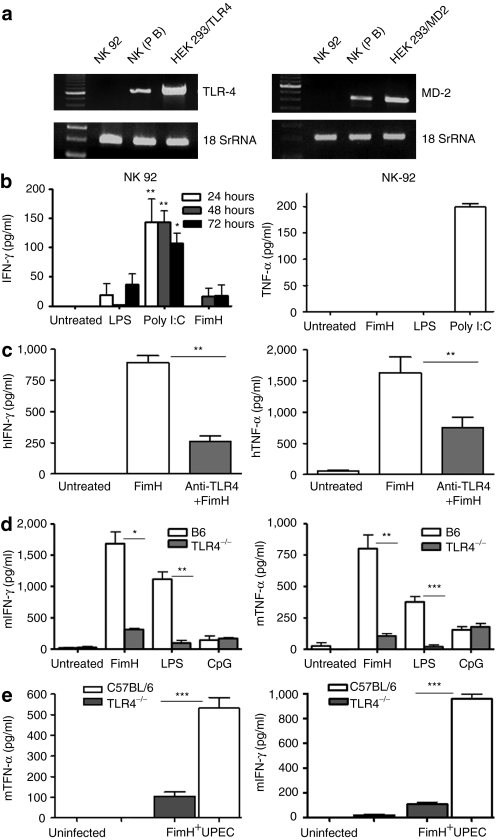
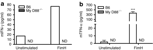
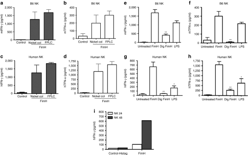
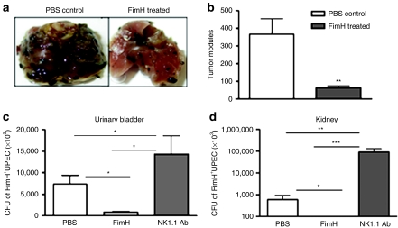
Although the importance of natural killer (NK) cells in innate immune responses against tumors or viral infections are well documented, their ability to directly recognize pathogens is less well defined. We have recently reported FimH, a bacterial fimbrial protein, as a novel Toll-like receptor (TLR)4 ligand that potently induces antiviral responses. Here, we investigated whether FimH either directly or indirectly can activate human and murine NK cells. We demonstrate that FimH potently activates both human and murine NK cells in vitro to induce cytokines [interferon (IFN)-gamma and tumor necrosis factor (TNF)-alpha] and cytotoxicity. Importantly, NK cells directly recognize FimH-expressing pathogens as FimH(+), but not FimH(-), bacteria were able to activate human NK cells. FimH activation of NK cells required TLR4 and MyD88 signaling, as NK cells from both TLR4(-/-) and MyD88(-/-) mice as well as human NK-92 cells, which lack TLR4, were all unresponsive to FimH. In addition, TLR4 neutralization significantly abrogated the response of human NK cells to FimH. Activation of purified NK cells by FimH was independent of lipopolysaccharide (LPS) or other bacterial contaminations. These data demonstrate for the first time that highly purified NK cells directly recognize and respond to FimH via TLR4-MyD88 pathways to aid innate protection against cancer or microbial infections.


Figures

Figure 1
Figure 1
FimH directly activates human and murine NK cell cytokine release and cytotoxicity. Human peripheral blood NK cells were cultured in the absence or presence of FimH (10 µg/ml), LPS (100 ng/ml), or CpG (10 µg/ml) for up to 72 hours and then cell-free supernatants harvested and analyzed for (a) IFN-γ and (b) TNF-α by specific enzyme-linked immunosorbent assay (ELISA). (Data are mean ± SEM of five separate experiments using NK cells from five donors cultured in triplicate wells.) Splenic NK cells from C57BL/6 mice were unstimulated or stimulated with FimH (10 µg/ml), LPS (100 ng/ml), or CpG (10 µg/ml) for 72 hours, cell-free supernatants harvested, and (c) IFN-γ or (d) TNF-α concentrations measured by specific ELISA. (Data are mean ± SEM of three individual experiments using NK cells from each of three mice cultured in triplicate wells.) (e) Human peripheral blood NK cells were cultured with or without FimH (10 µg/ml), LPS (100 ng/ml), or CpG (10 µg/ml) stimulations for 48 hours, co-cultured with chromium-labeled target K562 cells in triplicate wells at two different effector to target (E:T) ratios, 10:1 and 5:1, for 4 hours and were analyzed for cytotoxicity. (Data are mean ± SEM, n = 3.) (f) Human NK cells were untreated or treated with varying concentrations of FimH or CpG as shown in the figure for 48 hours and IFN-γ levels were measured in cell-free supernatants by ELISA. Data are mean ± SD from at least two separate experiments using two different volunteers. *P < 0.05, **P < 0.01, ***P < 0.001, comparisons were shown between FimH treatments and untreated controls and/or FimH treatments and LPS or CpG treatments. h, human; IFN, interferon; LPS, lipopolysaccharide; m, murine; NK, natural killer; TNF, tumor necrosis factor.
Figure 2
Figure 2
FimH activation of NK cells is direct. (a) Total splenocytes from RAG2−/− (n = 5) and RAG2−/−γc−/− (n = 3) mice were cultured with or without FimH (10 µg/ml) stimulations for 48 hours, and cell-free supernatants were assessed for IFN-γ production by enzyme-linked immunosorbent assay (ELISA). (b) Human peripheral blood mononuclear cells (PBMCs) and purified NK cells were cultured in absence or presence of FimH (10 µg/ml) for 24 hours and cell-free supernatants assessed for IL-12p40 secretions by ELISA (n = 3, mean ± SD). (c) Human peripheral blood NK cells initially purified by EasySep negative selection NK enrichment kit were further sorted by flow cytometry (98.8% pure NK cells) and cultured in triplicate wells with or without FimH (10 µg/ml) or LPS (100 ng/ml) for 48 hours and cell-free supernatants were assayed for IFN-γ by ELISA. (d) EasySep negative selection purified followed by flow cytometry sorted NK cells or PBMCs (as positive control) were induced with FimH (10 µg/ml) for 24 hours and supernatants were measured by ELISA for IL-12p40 production. Data for (c) and (d) are mean ± SD from two separate experiments with two donors). (e) Purified human peripheral blood NK cells were untreated or stimulated with FimH (10 µg/ml) for the indicated time periods, cells were then transferred onto slides by Shandon cytospin 4, acetone fixed and stained for NF-κB (green) using Alxafluor-488-labeled secondary antibody or counterstained (nuclear staining) with propidium iodide (red). Images were gained using LSM 510 confocal microscope using ×63 objectives (insets are higher magnification images). Bar = 20 µm. h, human; IFN, interferon; IL, interleukin; LPS, lipopolysaccharide; NK, natural killer; m, murine.
Figure 3
Figure 3
NK cells directly recognize FimH+ UPEC pathogens and become activated. Human peripheral blood CD56+ NK cells were either uninfected or infected ex vivo with FimH+ UPEC (Nu14) or δFimH-UPEC (Nu14-1) bacteria at an multiplicity of infection of 20 as described in Materials and Methods for 16 hours, fluorescence-activated cell sorting stained for intracellular perforin or surface expression of CD69, known NK-cell activation markers and analyzed by flow cytometry. CD56+CD3 lymphocyte populations (NK cells) expressing (a) perforin or (b) CD69 were gated to show NK cell activation phenotypes. Data represent three independent and reproducible experiments. Human peripheral blood NK cells were uninfected or infected with FimH+ or δFimH-UPEC bacteria similarly as mentioned earlier for 24 hours and cell-free supernatants were assessed for (c) IFN-γ or (d) TNF-α release by enzyme-linked immunosorbent assay. (e) Human peripheral NK cells were co-cultured with FimH+ or FimH UPEC at NK:UPEC ratio of 1:20 for 2 hours and cells washed, lysed, and cells lysates were plated onto Luria–Bertani agar for overnight and bacterial colony-forming units (CFU) were counted. Data represent mean ± SEM of at least two independent experiments involving two different donors. **P < 0.01. IFN, interferon; NK, natural killer; TNF, tumor necrosis factor; UPEC, uropathogenic E. coli.
Figure 4
Figure 4
FimH activation of NK cells requires TLR4 signaling. (a) Total RNA was isolated from human peripheral blood purified NK cells, NK-92 cells and 293t expressing TLR4/MD-2 cells and assessed mRNA expressions for TLR4 and MD-2 by reverse transcription-PCR. (b) Human NK-92 cells were untreated or stimulated with FimH, LPS, and poly I:C for up to 72 hours and cell-free supernatants were measured for IFN-γ and TNF-α productions by specific enzyme-linked immunosorbent assay (ELISA). Data represent mean ± SEM of three separate experiments done in triplicate wells. (c) Human peripheral blood NK cells were untreated, preincubated with anti-TLR4 mAb for 1 hour and/or treated with FimH (10 µg/ml), cell-free supernatants were harvested after 24 and 48 hours and analyzed for TNF-α and IFN-γ concentrations, respectively (n = 3, mean ± SEM). (d) Purified splenic NK cells from B6 or TLR4−/− mice were either untreated or treated with FimH (10 µg/ml), LPS (100 ng/ml), or CpG (10 µg/ml) and cell-free supernatants assessed for IFN-γ and TNF-α productions by ELISA (n = 5, mean ± SEM). (e) Purified splenic NK cells from B6 or TLR4−/− mice were co-cultured with FimH+ UPEC bacteria similarly as mentioned earlier for 24 hours and cell free supernatants were assessed for TNF-α or IFN-γ production by ELISA (data are mean ± SD). Experiments were repeated two times. *P < 0.05, **P < 0.01, ***P < 0.001. IFN, interferon; LPS, lipopolysaccharide; NK, natural killer; TLR4, Toll-like receptor 4; TNF, tumor necrosis factor; UPEC, uropathogenic E. coli.
Figure 5
Figure 5
NK cells activation by FimH depends on MyD88 signaling. Total splenocytes from C57BL/6 (n = 3) or MyD88−/− (n = 5) mice were isolated and cells were either left untreated or stimulated with FimH (10 µg/ml) for 24 hours for TNF-α and 48 hours for IFN-γ production. Cell-free supernatants were then harvested and analyzed by enzyme-linked immunosorbent assay for (a) IFN-γ and (b) TNF-α measurements (data are mean ± SEM of three repeated experiments). ***P < 0.001. ND, not detected; NK, natural killer; IFN, interferon; TNF, tumor necrosis factor.
Figure 6
Figure 6
FimH-stimulated NK cell activation is free of LPS contamination. (a,b) Purified splenic NK cells from C57BL/6 mice (n = 3), and (c,d) peripheral human NK cells (n = 3) were untreated or treated with column- and/or FPLC-purified recombinant FimH (10 µg/ml) for 24 and 48 hours, and cell-free supernatants were assessed by specific enzyme-linked immunosorbent assay (ELISA) for murine or human IFN-γ and TNF-α productions, respectively (data are mean ± SEM of three independent experiments). (e,f) Purified NK cells from C57BL/6 mice (n = 3) or from (g,h) human peripheral blood (n = 3) were cultured alone or in presence of FimH (10 µg/ml), proteinase-K/trypsin degraded FimH (10 µg/ml), and 100 ng/ml of LPS, cell-free supernatants harvested after 24 and 48 hours were then assessed by ELISA for TNF-α and IFN-γ, respectively (data are mean ± SEM of three separate experiments). (i) purified human peripheral NK cells were stimulated with recombinant vector His-tagged or recombinant (His-tagged) FimH protein for 48 hours and cell-free supernatants obtained at 24 and 48 hours were assayed for IFN-γ production by ELISA. Representative data from one donor is shown. *P < 0.05, **P < 0.01. FPLC, fast protein liquid chromatography; IFN, interferon; LPS, lipopolysaccharide; NK, natural killer; TNF, tumor necrosis factor.
Figure 7
Figure 7
FimH-induced protection against tumor and UPEC urinary tract infection are mediated by NK cells. (a) C57BL/6 mice were injected through foot pad in the hindlimb with PBS or FimH (50 µg). Forty-eight hours later, mice were challenged with 5 × 10⁵ B16F10 melanoma cells intravenously. (b) Lungs were harvested after 2 weeks and tumor nodules in the lungs were photographed and enumerated (n = 5–6/group, data are mean ± SEM). (c,d) B6 mice were injected with PBS or 50 µg of FimH on day 1 preinfection or with anti-NK1.1 antibody (200 µg) on days 2 and 1 preinfection. Twenty-four hours later, 108 CFU of FimH+UPEC (strain Nu14) bacteria were delivered into the urinary bladder through a soft catheter. Twenty-four hours later (c) urinary bladder and (d) kidney were removed, homogenized, plated onto LB agar and bacterial CFU enumerated. (n = 5–6, data are mean ± SEM). *P < 0.05, **P < 0.01, ***P < 0.001. LB, Luria–Bertani; CFU, colony-forming units; NK, natural killer; PBS, phosphate-buffered saline; TNF, tumor necrosis factor; UPEC, uropathogenic E. coli.

Similar articles

Cited by

References

    1. Biron CA, Nguyen KB, Pien GC, Cousens LP., and, Salazar-Mather TP. Natural killer cells in antiviral defense: function and regulation by innate cytokines. Annu Rev Immunol. 1999;17:189–220. - PubMed
    1. Wu J., and, Lanier LL. Natural killer cells and cancer. Adv Cancer Res. 2003;90:127–156. - PubMed
    1. Chalifour A, Jeannin P, Gauchat JF, Blaecke A, Malissard M, N'Guyen T, et al. Direct bacterial protein PAMP recognition by human NK cells involves TLRs and triggers α-defensin production. Blood. 2004;104:1778–1783. - PubMed
    1. Raulet DH. Interplay of natural killer cells and their receptors with the adaptive immune response. Nat Immunol. 2004;5:996–1002. - PubMed
    1. Biron CA., and, Brossay L. NK cells and NKT cells in innate defense against viral infections. Curr Opin Immunol. 2001;13:458–464. - PubMed

Publication types

LinkOut - more resources